
")

The characterization of aging is of great importance for social well-being and for the devastating impact of some neurological diseases associated with aging. Functional magnetic resonance imaging (fMRI) is an investigation tool capable of capturing the deterioration of the network functionality of the human brain associated with aging and neurodegeneration. However, fMRI suffers from limitations due to the variation of physiological noise with age , which is a confounding factor that is difficult to separate from the effect of neuronal origin. This project aims to address this scientific problem, which has important industrial implications in the health sector and biomedical industry, by characterizing the physiological origin of fMRI noise in aging and in the early stages of neurodegeneration in terms of changes of vascular reactivity and of micro-motion. We will also develop appropriate mitigation methods.
The project was funded by the Lazio Regional Government under POR FESR LAZIO 2014 - 2020 A0375-2020-36648, POR A0375E0085 grant (€ 149089.39).
Minimizing the effects of noise is a major challenge in functional magnetic resonance imaging (fMRI) in order to accurately recover the signal originating from neural activity. Higher spatial resolutions are possible at ultra-high (UH) magnetic fields (7T and above) than at more typical magnitudes. Nevertheless, UH-fMRI is also more susceptible to physiological and thermal noise. The best technique for denoising UH-fMRI data is still up for debate.
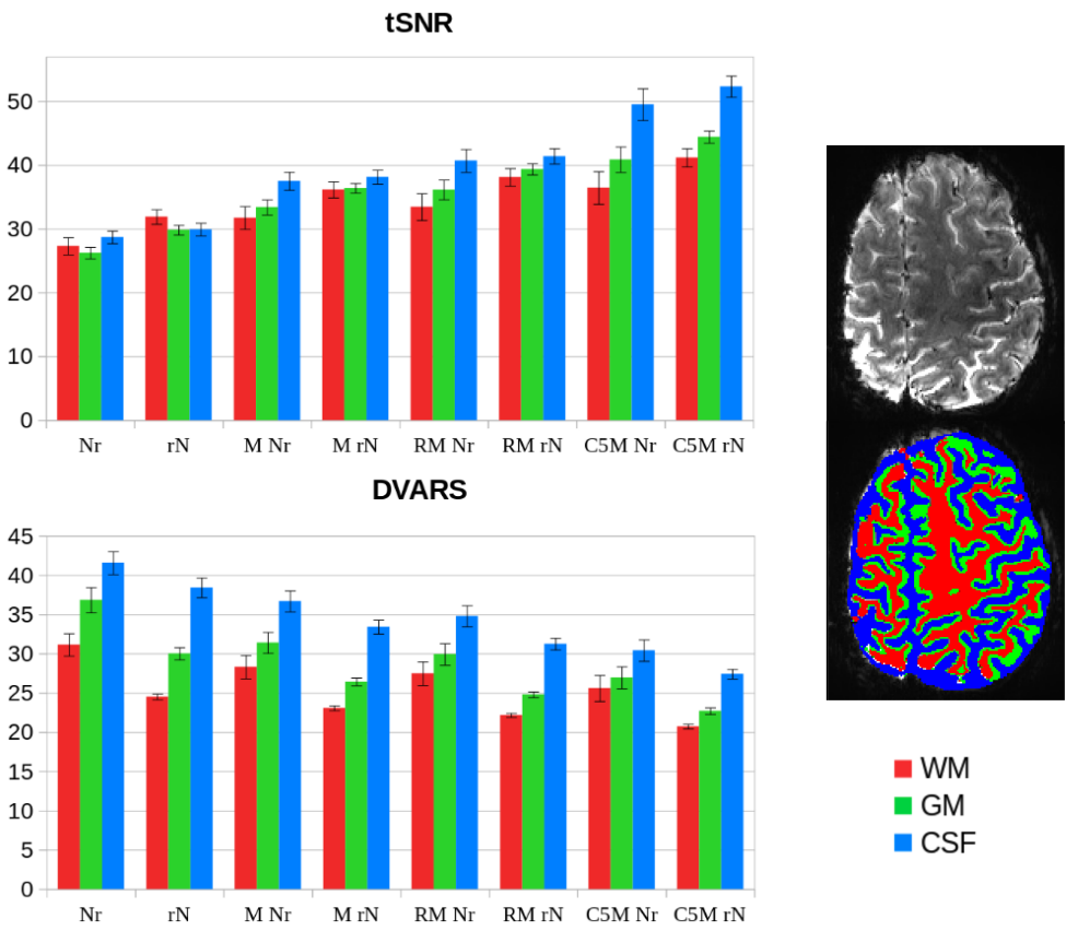
Using 7T resting-state fMRI data collected from seven healthy participants (three runs each), together with physiological (cardiac and respiratory) recordings, we evaluated the efficacy of few denoising pipelines based on recent developed algorithms (such as NORDIC [N], RETROICOR [R], and aCompCor [C5]). Each pipeline was created by permuting the order of the following analysis steps: motion correction, thermal denoising, and physiological denoising. Two quality metrics were computed to assess their efficacy: delta variation signal (DVARS) and temporal signal-to-noise ratio (tSNR). Additionally, we assessed the spatial distribution of physiological noise throughout the brain and its reproducibility across fMRI runs.
Compared to unprocessed fMRI data, all examined denoising methods enhanced the evaluated quality metrics, albeit to varying degrees, depending on the approach and the brain tissues taken into account. The figure shows the tSNR and DVARS values of the denoised datasets, averaged across all subjects and all fMRI runs, in the different tissues (white matter, WM, gray matter, GM, and cerebrospinal fluid, CSF). The analysis showed that applying thermal denoising (N) as the last step resulted in almost no improvement for all tissues. Conversely, performing N as the very first step consistently improved the quality metrics in all pipelines, as shown in figure, mainly in GM and WM. Finally, the quality ratings were enhanced by integrating physiological denoising as well, more so with aCompCor (C5MrN) than RETROICOR (RMrN). The aCompCor pipeline (C5MrN) achieved the best tSNR and DVARS values overall, with the greatest improvements for CSF, followed by GM and WM.
Regarding the spatial distribution of physiological noise, our results showed that the most affected brain tissue was the CSF, which also presented the highest level of repeatability among runs. These findings could help improve region-of-interest-based denoising algorithms (like aCompCor) by making it easier to identify the most dependable source of noise to exclude from the fMRI signal, making the neural activity detection more reliable.
Mauro DiNuzzo, Silvia Mangia and Federico Giove.
Manipulations of sleep-like slow-wave activity by noninvasive brain stimulation. Journal of Neuroscience Research 100:1218–1225.
Abstract Sleep is a universal and evolutionarily conserved behavior among many animal species, yet we do not have a fundamental understanding of why animals need to sleep. What we do know, however, is that sleep is critical for behavioral performance during the waking period and for long-term brain health. Here we provide an overview of some putative mechanisms that mediate the restorative effects of sleep, namely metabolic biosynthesis, fluid perfusion, and synaptic homeostasis. We then review recent experimental findings that advance the possibility of inducing sleep-like slow-wave activity (SWA) during wakefulness or enhance SWA during sleep in a top-down manner using noninvasive brain stimulation. SWA induction and SWA enhancement are believed to recapitulate the beneficial effects of sleep independent of the actual state of the subjects. If confirmed, these observations will change the way in which we investigate the neural correlates of sleep, thus paving the way for comprehending and actively controlling its restorative function.
DOI
Mauro DiNuzzo, Silvia Mangia, Marta Moraschi, Daniele Mascali, Gisela E Hagberg and Federico Giove.
Perception is associated with the brain's metabolic response to sensory stimulation. eLife 11.
Abstract Processing of incoming sensory stimulation triggers an increase of cerebral perfusion and blood oxygenation (neurovascular response) as well as an alteration of the metabolic neurochemical profile (neurometabolic response). Here we show in human primary visual cortex (V1) that perceived and unperceived isoluminant chromatic flickering stimuli designed to have similar neurovascular responses as measured by blood oxygenation level dependent functional MRI (BOLD-fMRI) have markedly different neurometabolic responses as measured by functional MRS. In particular, a significant regional buildup of lactate, an index of aerobic glycolysis, and glutamate, an index of malate-aspartate shuttle, occurred in V1 only when the flickering was perceived, without any relation with behavioral or physiological variables. Whereas the BOLD-fMRI signal in V1, a proxy for input to V1, was insensitive to flickering perception by design, the BOLD-fMRI signal in secondary visual areas was larger during perceived than unperceived flickering, indicating increased output from V1. These results demonstrate that the upregulation of energy metabolism induced by visual stimulation depends on the type of information processing taking place in V1, and that 1H-fMRS provides unique information about local input/output balance that is not measured by BOLD fMRI.
URL, DOI
Douglas L Rothman, Gerald A Dienel, Kevin L Behar, Fahmeed Hyder, Mauro DiNuzzo, Federico Giove and Silvia Mangia.
Glucose sparing by glycogenolysis (GSG) determines the relationship between brain metabolism and neurotransmission. Journal of Cerebral Blood Flow and Metabolism (42):844–860.
Abstract Over the last two decades, it has been established that glucose metabolic fluxes in neurons and astrocytes are proportional to the rates of the glutamate/GABA-glutamine neurotransmitter cycles in close to 1:1 stoichiometries across a wide range of functional energy demands. However, there is presently no mechanistic explanation for these relationships. We present here a theoretical meta-analysis that tests whether the brain’s unique compartmentation of glycogen metabolism in the astrocyte and the requirement for neuronal glucose homeostasis lead to the observed stoichiometries. We found that blood-brain barrier glucose transport can be limiting during activation and that the energy demand could only be met if glycogenolysis supports neuronal glucose metabolism by replacing the glucose consumed by astrocytes, a mechanism we call Glucose Sparing by Glycogenolysis (GSG). The predictions of the GSG model are in excellent agreement with a wide range of experimental results from rats, mice, tree shrews, and humans, which were previously unexplained. Glycogenolysis and glucose sparing dictate the energy available to support neuronal activity, thus playing a fundamental role in brain function in health and disease.
DOI
Julien Cohen-Adad, Eva Alonso-Ortiz, Mihael Abramovic, Carina Arneitz, Nicole Atcheson, Laura Barlow, Robert L Barry, Markus Barth, Marco Battiston, Christian Büchel, Matthew Budde, Virginie Callot, Anna J E Combes, Benjamin De Leener, Maxime Descoteaux, Paulo Loureiro Sousa, Marek Dostál, Julien Doyon, Adam Dvorak, Falk Eippert, Karla R Epperson, Kevin S Epperson, Patrick Freund, Jürgen Finsterbusch, Alexandru Foias, Michela Fratini, Issei Fukunaga, Claudia Gandini A M Wheeler-Kingshott, Giancarlo Germani, Guillaume Gilbert, Federico Giove, Charley Gros, Francesco Grussu, Akifumi Hagiwara, Pierre-Gilles Henry, Tomáš Horák, Masaaki Hori, James Joers, Kouhei Kamiya, Haleh Karbasforoushan, Miloš Keřkovský, Ali Khatibi, Joo-Won Kim, Nawal Kinany, Hagen Kitzler, Shannon Kolind, Yazhuo Kong, Petr Kudlička, Paul Kuntke, Nyoman D Kurniawan, Slawomir Kusmia, René Labounek, Maria Marcella Laganà, Cornelia Laule, Christine S Law, Christophe Lenglet, Tobias Leutritz, Yaou Liu, Sara Llufriu, Sean Mackey, Eloy Martinez-Heras, Loan Mattera, Igor Nestrasil, Kristin P O'Grady, Nico Papinutto, Daniel Papp, Deborah Pareto, Todd B Parrish, Anna Pichiecchio, Ferran Prados, Àlex Rovira, Marc J Ruitenberg, Rebecca S Samson, Giovanni Savini, Maryam Seif, Alan C Seifert, Alex K Smith, Seth A Smith, Zachary A Smith, Elisabeth Solana, Yuichi Suzuki, George Tackley, Alexandra Tinnermann, Jan Valošek, Dimitri Van De Ville, Marios C Yiannakas, Kenneth A Weber, Nikolaus Weiskopf, Richard G Wise, Patrik O Wyss and Junqian Xu.
Generic acquisition protocol for quantitative MRI of the spinal cord.. Nature protocols 16:4611–4632.
Abstract Quantitative spinal cord (SC) magnetic resonance imaging (MRI) presents many challenges, including a lack of standardized imaging protocols. Here we present a prospectively harmonized quantitative MRI protocol, which we refer to as the spine generic protocol, for users of 3T MRI systems from the three main manufacturers: GE, Philips and Siemens. The protocol provides guidance for assessing SC macrostructural and microstructural integrity: T1-weighted and T2-weighted imaging for SC cross-sectional area computation, multi-echo gradient echo for gray matter cross-sectional area, and magnetization transfer and diffusion weighted imaging for assessing white matter microstructure. In a companion paper from the same authors, the spine generic protocol was used to acquire data across 42 centers in 260 healthy subjects. The key details of the spine generic protocol are also available in an open-access document that can be found at https://github.com/spine-generic/protocols . The protocol will serve as a starting point for researchers and clinicians implementing new SC imaging initiatives so that, in the future, inclusion of the SC in neuroimaging protocols will be more common. The protocol could be implemented by any trained MR technician or by a researcher/clinician familiar with MRI acquisition.
DOI
Julien Cohen-Adad, Eva Alonso-Ortiz, Mihael Abramovic, Carina Arneitz, Nicole Atcheson, Laura Barlow, Robert L Barry, Markus Barth, Marco Battiston, Christian Büchel, Matthew Budde, Virginie Callot, Anna J E Combes, Benjamin De Leener, Maxime Descoteaux, Paulo Loureiro Sousa, Marek Dostál, Julien Doyon, Adam Dvorak, Falk Eippert, Karla R Epperson, Kevin S Epperson, Patrick Freund, Jürgen Finsterbusch, Alexandru Foias, Michela Fratini, Issei Fukunaga, Claudia A M Gandini Wheeler-Kingshott, Giancarlo Germani, Guillaume Gilbert, Federico Giove, Charley Gros, Francesco Grussu, Akifumi Hagiwara, Pierre-Gilles Henry, Tomáš Horák, Masaaki Hori, James Joers, Kouhei Kamiya, Haleh Karbasforoushan, Miloš Keřkovský, Ali Khatibi, Joo-Won Kim, Nawal Kinany, Hagen H Kitzler, Shannon Kolind, Yazhuo Kong, Petr Kudlička, Paul Kuntke, Nyoman D Kurniawan, Slawomir Kusmia, René Labounek, Maria Marcella Laganà, Cornelia Laule, Christine S Law, Christophe Lenglet, Tobias Leutritz, Yaou Liu, Sara Llufriu, Sean Mackey, Eloy Martinez-Heras, Loan Mattera, Igor Nestrasil, Kristin P O'Grady, Nico Papinutto, Daniel Papp, Deborah Pareto, Todd B Parrish, Anna Pichiecchio, Ferran Prados, Àlex Rovira, Marc J Ruitenberg, Rebecca S Samson, Giovanni Savini, Maryam Seif, Alan C Seifert, Alex K Smith, Seth A Smith, Zachary A Smith, Elisabeth Solana, Y Suzuki, George Tackley, Alexandra Tinnermann, Jan Valošek, Dimitri Van De Ville, Marios C Yiannakas, Kenneth A Weber Ii, Nikolaus Weiskopf, Richard G Wise, Patrik O Wyss and Junqian Xu.
Open-access quantitative MRI data of the spinal cord and reproducibility across participants, sites and manufacturers.. Scientific data 8:219.
Abstract In a companion paper by Cohen-Adad et al. we introduce the spine generic quantitative MRI protocol that provides valuable metrics for assessing spinal cord macrostructural and microstructural integrity. This protocol was used to acquire a single subject dataset across 19 centers and a multi-subject dataset across 42 centers (for a total of 260 participants), spanning the three main MRI manufacturers: GE, Philips and Siemens. Both datasets are publicly available via git-annex. Data were analysed using the Spinal Cord Toolbox to produce normative values as well as inter/intra-site and inter/intra-manufacturer statistics. Reproducibility for the spine generic protocol was high across sites and manufacturers, with an average inter-site coefficient of variation of less than 5% for all the metrics. Full documentation and results can be found at https://spine-generic.rtfd.io/. The datasets and analysis pipeline will help pave the way towards accessible and reproducible quantitative MRI in the spinal cord.
DOI
Riccardo De Feo, Artem Shatilo, Alejandra Sierra, Juan-Miguel Valverde, Olli Gröhn, Federico Giove and Jussi Tohka.
Automated joint skull-stripping and segmentation with Multi-Task U-Net in large mouse brain MRI databases. Neuroimage 229:117734.
Abstract Skull-stripping and region segmentation are fundamental steps in preclinical magnetic resonance imaging (MRI) studies, and these common procedures are usually performed manually. We present Multi-task U-Net (MU-Net), a convolutional neural network designed to accomplish both tasks simultaneously. MU-Net achieved higher segmentation accuracy than state-of-the-art multi-atlas segmentation methods with an inference time of 0.35 s and no pre-processing requirements. We trained and validated MU-Net on 128 T2-weighted mouse MRI volumes as well as on the publicly available MRM NeAT dataset of 10 MRI volumes. We tested MU-Net with an unusually large dataset combining several independent studies consisting of 1782 mouse brain MRI volumes of both healthy and Huntington animals, and measured average Dice scores of 0.906 (striati), 0.937 (cortex), and 0.978 (brain mask). Further, we explored the effectiveness of our network in the presence of different architectural features, including skip connections and recently proposed framing connections, and the effects of the age range of the training set animals. These high evaluation scores demonstrate that MU-Net is a powerful tool for segmentation and skull-stripping, decreasing inter and intra-rater variability of manual segmentation. The MU-Net code and the trained model are publicly available at https://github.com/Hierakonpolis/MU-Net.
DOI
Daniele Mascali, Marta Moraschi, Mauro DiNuzzo, Silvia Tommasin, Michela Fratini, Tommaso Gili, Richard G Wise, Silvia Mangia, Emiliano Macaluso and Federico Giove.
Evaluation of denoising strategies for task-based functional connectivity: Equalizing residual motion artifacts between rest and cognitively demanding tasks. Human Brain Mapping 42:1805–1828.
Abstract In-scanner head motion represents a major confounding factor in functional connectivity studies and it raises particular concerns when motion correlates with the effect of interest. One such instance regards research focused on functional connectivity modulations induced by sustained cognitively demanding tasks. Indeed, cognitive engagement is generally associated with substantially lower in-scanner movement compared with unconstrained, or minimally constrained, conditions. Consequently, the reliability of condition-dependent changes in functional connectivity relies on effective denoising strategies. In this study, we evaluated the ability of common denoising pipelines to minimize and balance residual motion-related artifacts between resting-state and task conditions. Denoising pipelines—including realignment/tissue-based regression, PCA/ICA-based methods (aCompCor and ICA-AROMA, respectively), global signal regression, and censoring of motion-contaminated volumes—were evaluated according to a set of benchmarks designed to assess either residual artifacts or network identifiability. We found a marked heterogeneity in pipeline performance, with many approaches showing a differential efficacy between rest and task conditions. The most effective approaches included aCompCor, optimized to increase the noise prediction power of the extracted confounding signals, and global signal regression, although both strategies performed poorly in mitigating the spurious distance-dependent association between motion and connectivity. Censoring was the only approach that substantially reduced distance-dependent artifacts, yet this came at the great cost of reduced network identifiability. The implications of these findings for best practice in denoising task-based functional connectivity data, and more generally for resting-state data, are discussed.
DOI
Paolo Miocchi, Alejandra Sierra, Laura Maugeri, Eleonora Stefanutti, Ali Abdollahzadeh, Fabio Mangini, Marta Moraschi, Inna Bukreeva, Lorenzo Massimi, Francesco Brun, Jussi Tohka, Olli Gröhn, Alberto Mittone, Alberto Bravin, Charles Nicaise, Federico Giove, Alessia Cedola and Michela Fratini.
Steerable3D: an ImageJ plugin for neurovascular enhancement in 3-D segmentation. Physica Medica 81:197–209.
DOI
Michela Fratini, Ali Abdollahzadeh, Mauro DiNuzzo, Raimo A Salo, Laura Maugeri, Alessia Cedola, Federico Giove, Olli Gröhn, Jussi Tohka and Alejandra Sierra.
Multiscale imaging approach for studying the central nervous system: methodology and perspective. Frontiers in Neuroscience 14:72.
Abstract Non-invasive imaging methods have become essential tools for understanding the central nervous system (CNS) in health and disease. In particular, magnetic resonance imaging (MRI) techniques provide information about the anatomy, microstructure, and function of the brain and spinal cord in vivo non-invasively. However, MRI is limited by its spatial resolution and signal specificity. In order to mitigate these shortcomings, it is crucial to validate MRI with an array of ancillary ex vivo imaging techniques. These techniques include histological methods, such as light and electron microscopy, which can provide specific information on the tissue structure in healthy and diseased brain and spinal cord, at cellular and subcellular level. However, these conventional histological techniques are intrinsically 2-dimensional (2D) and, as a result of sectioning, lack volumetric information of the tissue. This limitation can be overcome with genuine three-dimensional (3D) imaging approaches of the tissue. 3D highly resolved information of the CNS achievable by means of other imaging techniques can complement and improve the interpretation of MRI measurements. In this article, we provide an overview of different 3D imaging techniques that can be used to validate MRI. As an example, we introduce an approach of how to combine diffusion MRI and synchrotron X-ray phase contrast tomography (SXRPCT) data. Our approach paves the way for a new multiscale assessment of the CNS allowing to validate and to improve our understanding of in vivo imaging (such as MRI).
DOI
Marta Moraschi, Daniele Mascali, Silvia Tommsain, Tommaso Gili, Ibrahim Eid Hassan, Michela Fratini, Mauro DiNuzzo, Richard G Wise, Silvia Mangia, Emiliano Macaluso and Federico Giove.
Brain Network Modularity During a Sustained Working-Memory Task. Frontiers in Physiology 11:422.
Abstract Spontaneous oscillations of the blood oxygenation level-dependent (BOLD) signal are spatially synchronized within specific brain networks and are thought to reflect synchronized brain activity. Networks are modulated by the performance of a task, even if the exact features and degree of such modulations are still elusive. The presence of networks showing anticorrelated fluctuations lend initially to suppose that a competitive relationship between the default mode network (DMN) and task positive networks (TPNs) supports the efficiency of brain processing. However, more recent results indicate that cooperative and competitive dynamics between networks coexist during task performance. In this study, we used graph analysis to assess the functional relevance of the topological reorganization of brain networks ensuing the execution of a steady state working-memory (WM) task. Our results indicate that the performance of an auditory WM task is associated with a switching between different topological configurations of several regions of specific networks, including frontoparietal, ventral attention, and dorsal attention areas, suggesting segregation of ventral attention regions in the presence of increased overall integration. However, the correct execution of the task requires integration between components belonging to all the involved networks.
DOI
Riccardo De Feo and Federico Giove.
Towards an efficient segmentation of small rodents brain: a short critical review. Journal of Neuroscience Methods 323:82–89.
Abstract One of the most common tasks in small rodents MRI pipelines is the voxel-wise segmentation of the volume in multiple classes. While many segmentation schemes have been developed for the human brain, fewer are available for rodent MRI, often by adaptation from human neuroimaging. Common methods include atlas-based and clustering schemes. The former labels the target volume by registering one or more pre-labeled atlases using a deformable registration method, in which case the result depends on the quality of the reference volumes, the registration algorithm and the label fusion approach, if more than one atlas is employed. The latter is based on an expectation maximization procedure to maximize the variance between voxel categories, and is often combined with Markov Random Fields and the atlas based approach to include spatial information, priors, and improve the classification accuracy. Our primary goal is to critically review the state of the art of rat and mouse segmentation of neuro MRI volumes and compare the available literature on popular, readily and freely available MRI toolsets, including SPM, FSL and ANTs, when applied to this task in the context of common pre-processing steps. Furthermore, we will briefly address the emerging Deep Learning methods for the segmentation of medical imaging, and the perspectives for applications to small rodents.
DOI
Mauro DiNuzzo, Daniele Mascali, Marta Moraschi, Giorgia Bussu, Laura Maugeri, Fabio Mangini, Michela Fratini and Federico Giove.
Brain networks underlying eye’s pupil dynamics. Frontiers in Neuroscience 13:965.
Abstract Phasic changes in eye’s pupil diameter have been repeatedly observed during cognitive, emotional and behavioral activity in mammals. Although pupil diameter is known to be associated with noradrenergic firing in the pontine Locus Coeruleus (LC), thus far the causal chain coupling spontaneous pupil dynamics to specific cortical brain networks remains unknown. In the present study, we acquired steady-state blood oxygenation level-dependent (BOLD) functional magnetic resonance imaging (fMRI) data combined with eye-tracking pupillometry from fifteen healthy subjects that were trained to maintain a constant attentional load. Regression analysis revealed widespread visual and sensorimotor BOLD-fMRI deactivations correlated with pupil diameter. Furthermore, we found BOLD-fMRI activations correlated with pupil diameter change rate within a set of brain regions known to be implicated in selective attention, salience, error-detection and decision-making. These regions included LC, thalamus, posterior cingulate cortex (PCC), dorsal anterior cingulate and paracingulate cortex (dACC/PaCC), orbitofrontal cortex (OFC), and right anterior insular cortex (rAIC). Granger-causality analysis performed on these regions yielded a complex pattern of interdependence, wherein LC and pupil dynamics were far apart in the network and separated by several cortical stages. Functional connectivity (FC) analysis revealed the ubiquitous presence of the superior frontal gyrus (SFG) in the networks identified by the brain regions correlated to the pupil diameter change rate. No significant correlations were observed between pupil dynamics, regional activation and behavioral performance. Based on the involved brain regions, we speculate that pupil dynamics reflects brain processing implicated in changes between self- and environment-directed awareness.
DOI
Fabio Mangini, Mauro DiNuzzo, Laura Maugeri, Marta Moraschi, Daniele Mascali, Alessia Cedola, Fabrizio Frezza, Federico Giove and Michela Fratini.
Numerical simulation of the Blood Oxygenation Level-Dependent functional magnetic resonance signal using finite element method. International Journal for Numerical Methods in Biomedical Engineering, pages e3290.
DOI
Petr Bednařík, Ivan Tkáč, Federico Giove, Lynn E Eberly, Dinesh K Deelchand, Felipe R Barreto and Silvia Mangia.
Neurochemical responses to chromatic and achromatic stimuli in the human visual cortex.. Journal of cerebral blood flow and metabolism 38:347–359.
Abstract In the present study, we aimed at determining the metabolic responses of the human visual cortex during the presentation of chromatic and achromatic stimuli, known to preferentially activate two separate clusters of neuronal populations (called "blobs" and "interblobs") with distinct sensitivity to color or luminance features. Since blobs and interblobs have different cytochrome-oxidase (COX) content and micro-vascularization level (i.e., different capacities for glucose oxidation), different functional metabolic responses during chromatic vs. achromatic stimuli may be expected. The stimuli were optimized to evoke a similar load of neuronal activation as measured by the bold oxygenation level dependent (BOLD) contrast. Metabolic responses were assessed using functional 1H MRS at 7 T in 12 subjects. During both chromatic and achromatic stimuli, we observed the typical increases in glutamate and lactate concentration, and decreases in aspartate and glucose concentration, that are indicative of increased glucose oxidation. However, within the detection sensitivity limits, we did not observe any difference between metabolic responses elicited by chromatic and achromatic stimuli. We conclude that the higher energy demands of activated blobs and interblobs are supported by similar increases in oxidative metabolism despite the different capacities of these neuronal populations.
DOI
Daniele Mascali, Mauro DiNuzzo, Laura Serra, Silvia Mangia, Bruno Maraviglia, Marco Bozzali and Federico Giove.
Disruption of Semantic Network in Mild Alzheimer's Disease Revealed by Resting-State fMRI.. Neuroscience 371:38–48.
Abstract Subtle semantic deficits can be observed in Alzheimer's disease (AD) patients even in the early stages of the illness. In this work, we tested the hypothesis that the semantic control network is deregulated in mild AD patients. We assessed the integrity of the semantic control system using resting-state functional magnetic resonance imaging in a cohort of patients with mild AD (n = 38; mean mini-mental state examination = 20.5) and in a group of age-matched healthy controls (n = 19). Voxel-wise analysis spatially constrained in the left fronto-temporal semantic control network identified two regions with altered functional connectivity (FC) in AD patients, specifically in the pars opercularis (POp, BA44) and in the posterior middle temporal gyrus (pMTG, BA21). Using whole-brain seed-based analysis, we demonstrated that these two regions have altered FC even beyond the semantic control network. In particular, the pMTG displayed a wide-distributed pattern of lower connectivity to several brain regions involved in language-semantic processing, along with a possibly compensatory higher connectivity to the Wernicke's area. We conclude that in mild AD brain regions belonging to the semantic control network are abnormally connected not only within the network, but also to other areas known to be critical for language processing.
DOI
Laura Maugeri, Mauro DiNuzzo, Marta Moraschi, Charles Nicaise, Inna Bukreeva, Fabio Mangini, Federico Giove, Alessia Cedola and Michela Fratini.
Fractal dimension analysis of high-resolution X-ray phase contrast micro-tomography images at different threshold levels in a mouse spinal cord. Condensed Matter 3:48.
DOI
Laura Maugeri, Marta Moraschi, Paul E Summers, Stefania Favilla, Carlo Adolfo Porro, Alessia Cedola, Eleonora Stefanutti, Paolo Miocchi, Federico Giove and Michela Fratini.
Assessing denoising strategies for fMRI in spinal cord and Brainstem. Journal of Instrumentation 13:C02028.
Abstract Functional Magnetic Resonance Imaging (fMRI) based on Blood Oxygenation Level Dependent (BOLD) contrast has become one of the most powerful tools in neuroscience research. On the other hand, fMRI approaches have seen limited use in the study of spinal cord and subcortical brain regions (such as the brainstem and portions of the diencephalon). Indeed obtaining good BOLD signal in these areas still represents a technical and scientific challenge, due to poor control of physiological noise and to a limited overall quality of the functional series. A solution can be found in the combination of optimized experimental procedures at acquisition stage, and well-adapted artifact mitigation procedures in the data processing. In this framework, we studied two different data processing strategies to reduce physiological noise in cortical and subcortical brain regions and in the spinal cord, based on the aCompCor and RETROICOR denoising tools respectively. The study, performed in healthy subjects, was carried out using an ad hoc isometric motor task. We observed an increased signal to noise ratio in the denoised functional time series in the spinal cord and in the subcortical brain region.
DOI
Andrea Romano, Marta Moraschi, Riccardo Cornia, Alessandro Bozzao, Maria Camilla Rossi-Espagnet, Federico Giove, Giorgio Albertini and Alberto Pierallini.
White matter involvement in young non-demented Down's syndrome subjects: a tract-based spatial statistic analysis.. Neuroradiology 60:1335–1341.
Abstract Cognitive decline in Down syndrome generally shows neurodegenerative aspects similar to what is observed in Alzheimer's disease. Few studies reported information on white matter integrity. The aim of this study was to evaluate white matter alterations in a cohort of young Down subjects, without dementia, by means of DTI technique, compared to a normal control group. The study group consisted of 17 right-handed subjects with DS and many control subjects. All individuals participating in this study were examined by MR exam including DTI acquisition (32 non-coplanar directions); image processing and analysis were performed using FMRIB Software Library (FSL version 4.1.9, http://www.fmrib.ox.ac.uk/fsl )) software package. Finally, the diffusion tensor was estimated voxel by voxel and the FA map derived from the tensor. A two-sample t test was performed to assess differences between DS and control subjects. The FA is decreased in DS subjects, compared to control subjects, in the region of the anterior thalamic radiation, the inferior fronto-occipital fasciculum, the inferior longitudinal fasciculum, and the cortico-spinal tract, bilaterally. The early white matter damage visible in our DS subjects could have great impact in the therapeutic management, in particular in better adapting the timing of therapies to counteract the toxic effect of the deposition of amyloid that leads to oxidative stress.
DOI
Eleonora Stefanutti, Alejandra Sierra, Paolo Miocchi, Lorenzo Massimi, Francesco Brun, Laura Maugeri, Inna Bukreeva, Annti Nurmi, Giovanni Begani Provinciali, Giuliana Tromba, Olli Gröhn, Federico Giove, Alessia Cedola and Michela Fratini.
Assessment of the effects of different sample perfusion procedures on phase-contrast tomographic images of mouse spinal cord. Journal of Instrumentation 13:C03027.
Abstract Synchrotron X-ray Phase Contrast micro-Tomography (SXrPCμT) is a powerful tool in the investigation of biological tissues, including the central nervous system (CNS), and it allows to simultaneously detect the vascular and neuronal network avoiding contrast agents or destructive sample preparations. However, specific sample preparation procedures aimed to optimize the achievable contrast- and signal-to-noise ratio (CNR and SNR, respectively) are required. Here we report and discuss the effects of perfusion with two different fixative agents (ethanol and paraformaldehyde) and with a widely used contrast medium (MICROFIL) on mouse spinal cord. As a main result, we found that ethanol enhances contrast at the grey/white matter interface and increases the contrast in correspondence of vascular features and fibres, thus providing an adequate spatial resolution to visualise the vascular network at the microscale. On the other hand, ethanol is known to induce tissue dehydration, likely reducing cell dimensions below the spatial resolution limit imposed by the experimental technique. Nonetheless, neurons remain well visible using either perfused paraformaldehyde or MICROFIL compound, as these latter media do not affect tissues with dehydration effects. Paraformaldehyde appears as the best compromise: it is not a contrast agent, like MICROFIL, but it is less invasive than ethanol and permits to visualise well both cells and blood vessels. However, a quantitative estimation of the relative grey matter volume of each sample has led us to conclude that no significant alterations in the grey matter extension compared to the white matter occur as a consequence of the perfusion procedures tested in this study.
DOI
Silvia Tommasin, Daniele Mascali, Marta Moraschi, Tommaso Gili, Ibrahim Eid Hassan, Michela Fratini, Mauro DiNuzzo, Richard G Wise, Silvia Mangia, Emiliano Macaluso and Federico Giove.
Scale-invariant rearrangement of resting state networks in the human brain under sustained stimulation.. NeuroImage 179:570–581.
Abstract Brain activity at rest is characterized by widely distributed and spatially specific patterns of synchronized low-frequency blood-oxygenation level-dependent (BOLD) fluctuations, which correspond to physiologically relevant brain networks. This network behaviour is known to persist also during task execution, yet the details underlying task-associated modulations of within- and between-network connectivity are largely unknown. In this study we exploited a multi-parametric and multi-scale approach to investigate how low-frequency fluctuations adapt to a sustained n-back working memory task. We found that the transition from the resting state to the task state involves a behaviourally relevant and scale-invariant modulation of synchronization patterns within both task-positive and default mode networks. Specifically, decreases of connectivity within networks are accompanied by increases of connectivity between networks. In spite of large and widespread changes of connectivity strength, the overall topology of brain networks is remarkably preserved. We show that these findings are strongly influenced by connectivity at rest, suggesting that the absolute change of connectivity (i.e., disregarding the baseline) may be not the most suitable metric to study dynamic modulations of functional connectivity. Our results indicate that a task can evoke scale-invariant, distributed changes of BOLD fluctuations, further confirming that low frequency BOLD oscillations show a specialized response and are tightly bound to task-evoked activation.
DOI
I Bukreeva, G Campi, Michela Fratini, R Spanò, D Bucci, G Battaglia, Federico Giove, A Bravin, A Uccelli, C Venturi, M Mastrogiacomo and A Cedola.
Quantitative 3D investigation of Neuronal network in mouse spinal cord model.. Scientific reports 7:41054.
Abstract The investigation of the neuronal network in mouse spinal cord models represents the basis for the research on neurodegenerative diseases. In this framework, the quantitative analysis of the single elements in different districts is a crucial task. However, conventional 3D imaging techniques do not have enough spatial resolution and contrast to allow for a quantitative investigation of the neuronal network. Exploiting the high coherence and the high flux of synchrotron sources, X-ray Phase-Contrast multiscale-Tomography allows for the 3D investigation of the neuronal microanatomy without any aggressive sample preparation or sectioning. We investigated healthy-mouse neuronal architecture by imaging the 3D distribution of the neuronal-network with a spatial resolution of 640 nm. The high quality of the obtained images enables a quantitative study of the neuronal structure on a subject-by-subject basis. We developed and applied a spatial statistical analysis on the motor neurons to obtain quantitative information on their 3D arrangement in the healthy-mice spinal cord. Then, we compared the obtained results with a mouse model of multiple sclerosis. Our approach paves the way to the creation of a "database" for the characterization of the neuronal network main features for a comparative investigation of neurodegenerative diseases and therapies.
DOI
Mauro DiNuzzo, Federico Giove, Bruno Maraviglia and Silvia Mangia.
Computational Flux Balance Analysis Predicts that Stimulation of Energy Metabolism in Astrocytes and their Metabolic Interactions with Neurons Depend on Uptake of K+ Rather than Glutamate.. Neurochemical research 42:202–216.
Abstract Brain activity involves essential functional and metabolic interactions between neurons and astrocytes. The importance of astrocytic functions to neuronal signaling is supported by many experiments reporting high rates of energy consumption and oxidative metabolism in these glial cells. In the brain, almost all energy is consumed by the Na+/K+ ATPase, which hydrolyzes 1 ATP to move 3 Na+ outside and 2 K+ inside the cells. Astrocytes are commonly thought to be primarily involved in transmitter glutamate cycling, a mechanism that however only accounts for few \ percent of brain energy utilization. In order to examine the participation of astrocytic energy metabolism in brain ion homeostasis, here we attempted to devise a simple stoichiometric relation linking glutamatergic neurotransmission to Na+ and K+ ionic currents. To this end, we took into account ion pumps and voltage/ligand-gated channels using the stoichiometry derived from available energy budget for neocortical signaling and incorporated this stoichiometric relation into a computational metabolic model of neuron-astrocyte interactions. We aimed at reproducing the experimental observations about rates of metabolic pathways obtained by 13C-NMR spectroscopy in rodent brain. When simulated data matched experiments as well as biophysical calculations, the stoichiometry for voltage/ligand-gated Na+ and K+ fluxes generated by neuronal activity was close to a 1:1 relationship, and specifically 63/58 Na+/K+ ions per glutamate released. We found that astrocytes are stimulated by the extracellular K+ exiting neurons in excess of the 3/2 Na+/K+ ratio underlying Na+/K+ ATPase-catalyzed reaction. Analysis of correlations between neuronal and astrocytic processes indicated that astrocytic K+ uptake, but not astrocytic Na+-coupled glutamate uptake, is instrumental for the establishment of neuron-astrocytic metabolic partnership. Our results emphasize the importance of K+ in stimulating the activation of astrocytes, which is relevant to the understanding of brain activity and energy metabolism at the cellular level.
DOI
Mauro DiNuzzo, Daniele Mascali, Marta Moraschi, Giorgia Bussu, Bruno Maraviglia, Silvia Mangia and Federico Giove.
Temporal Information Entropy of the Blood-Oxygenation Level-Dependent Signals Increases in the Activated Human Primary Visual Cortex.. Frontiers in physics 5:7.
Abstract Time-domain analysis of blood-oxygenation level-dependent (BOLD) signals allows the identification of clusters of voxels responding to photic stimulation in primary visual cortex (V1). However, the characterization of information encoding into temporal properties of the BOLD signals of an activated cluster is poorly investigated. Here, we used Shannon entropy to determine spatial and temporal information encoding in the BOLD signal within the most strongly activated area of the human visual cortex during a hemifield photic stimulation. We determined the distribution profile of BOLD signals during epochs at rest and under stimulation within small (19-121 voxels) clusters designed to include only voxels driven by the stimulus as highly and uniformly as possible. We found consistent and significant increases (2-4\ percent on average) in temporal information entropy during activation in contralateral but not ipsilateral V1, which was mirrored by an expected loss of spatial information entropy. These opposite changes coexisted with increases in both spatial and temporal mutual information (i.e., dependence) in contralateral V1. Thus, we showed that the first cortical stage of visual processing is characterized by a specific spatiotemporal rearrangement of intracluster BOLD responses. Our results indicate that while in the space domain BOLD maps may be incapable of capturing the functional specialization of small neuronal populations due to relatively low spatial resolution, some information encoding may still be revealed in the temporal domain by an increase of temporal information entropy.
DOI
Silvia Mangia, Alena Svatkova, Daniele Mascali, Mikko J Nissi, Philip C Burton, Petr Bednarik, Edward J Auerbach, Federico Giove, Lynn E Eberly, Michael J Howell, Igor Nestrasil, Paul J Tuite and Shalom Michaeli.
Multi-modal Brain MRI in Subjects with PD and iRBD.. Frontiers in neuroscience 11:709.
Abstract Idiopathic rapid eye movement sleep behavior disorder (iRBD) is a condition that often evolves into Parkinson's disease (PD). Therefore, by monitoring iRBD it is possible to track the neurodegeneration of individuals who may progress to PD. Here we aimed at piloting the characterization of brain tissue properties in mid-brain subcortical regions of 10 healthy subjects, 8 iRBD, and 9 early-diagnosed PD. We used a battery of magnetic resonance imaging (MRI) contrasts at 3 T, including adiabatic and non-adiabatic rotating frame techniques developed by our group, along with diffusion tensor imaging (DTI) and resting-state fMRI. Adiabatic T1ρ and T2ρ, and non-adiabatic RAFF4 (Relaxation Along a Fictitious Field in the rotating frame of rank 4) were found to have lower coefficient of variations and higher sensitivity to detect group differences as compared to DTI parameters such as fractional anisotropy and mean diffusivity. Significantly longer T1ρ were observed in the amygdala of PD subjects vs. controls, along with a trend of lower functional connectivity as measured by regional homogeneity, thereby supporting the notion that amygdalar dysfunction occurs in PD. Significant abnormalities in reward networks occurred in iRBD subjects, who manifested lower network strength of the accumbens. In agreement with previous studies, significantly longer T1ρ occurred in the substantia nigra compacta of PD vs. controls, indicative of neuronal degeneration, while regional homogeneity was lower in the substantia nigra reticulata. Finally, other trend-level findings were observed, i.e., lower RAFF4 and T2ρ in the midbrain of iRBD subjects vs. controls, possibly indicating changes in non-motor features as opposed to motor function in the iRBD group. We conclude that rotating frame relaxation methods along with functional connectivity measures are valuable to characterize iRBD and PD subjects, and with proper validation in larger cohorts may provide pathological signatures of iRBD and PD.
DOI
Silvia Tommasin, Daniele Mascali, Tommaso Gili, Ibrahim Eid Assan, Marta Moraschi, Michela Fratini, Richard G Wise, Emiliano Macaluso, Silvia Mangia and Federico Giove.
Task-Related Modulations of BOLD Low-Frequency Fluctuations within the Default Mode Network.. Frontiers in Physics 5(31).
Abstract Spontaneous low-frequency Blood-Oxygenation Level-Dependent (BOLD) signals acquired during resting state are characterized by spatial patterns of synchronous fluctuations, ultimately leading to the identification of robust brain networks. The resting-state brain networks, including the Default Mode Network (DMN), are demonstrated to persist during sustained task execution, but the exact features of task-related changes of network properties are still not well characterized. In this work we sought to examine in a group of 20 healthy volunteers (age 33 ± 6 years, 8 F/12 M) the relationship between changes of spectral and spatiotemporal features of one prominent resting-state network, namely the DMN, during the continuous execution of a working memory n-back task. We found that task execution impacted on both functional connectivity and amplitude of BOLD fluctuations within large parts of the DMN, but these changes correlated between each other only in a small area of the posterior cingulate. We conclude that combined analysis of multiple parameters related to connectivity, and their changes during the transition from resting state to continuous task execution, can contribute to a better understanding of how brain networks rearrange themselves in response to a task.
DOI
Petr Bednařík, Ivan Tkáč, Federico Giove, Mauro DiNuzzo, Dinesh K Deelchand, Uzay E Emir, Lynn E Eberly and Silvia Mangia.
Neurochemical and BOLD responses during neuronal activation measured in the human visual cortex at 7 Tesla.. Journal of Cerebral Blood Flow and Metabolism 35(4):601–610.
Abstract Several laboratories have consistently reported small concentration changes in lactate, glutamate, aspartate, and glucose in the human cortex during prolonged stimuli. However, whether such changes correlate with blood oxygenation level-dependent functional magnetic resonance imaging (BOLD-fMRI) signals have not been determined. The present study aimed at characterizing the relationship between metabolite concentrations and BOLD-fMRI signals during a block-designed paradigm of visual stimulation. Functional magnetic resonance spectroscopy (fMRS) and fMRI data were acquired from 12 volunteers. A short echo-time semi-LASER localization sequence optimized for 7 Tesla was used to achieve full signal-intensity MRS data. The group analysis confirmed that during stimulation lactate and glutamate increased by 0.26 ± 0.06 μmol/g (~30\ percent ) and 0.28 ± 0.03 μmol/g (~3\ percent ), respectively, while aspartate and glucose decreased by 0.20 ± 0.04 μmol/g (~5\ percent ) and 0.19 ± 0.03 μmol/g (~16\ percent ), respectively. The single-subject analysis revealed that BOLD-fMRI signals were positively correlated with glutamate and lactate concentration changes. The results show a linear relationship between metabolic and BOLD responses in the presence of strong excitatory sensory inputs, and support the notion that increased functional energy demands are sustained by oxidative metabolism. In addition, BOLD signals were inversely correlated with baseline γ-aminobutyric acid concentration. Finally, we discussed the critical importance of taking into account linewidth effects on metabolite quantification in fMRS paradigms.
DOI
Mauro DiNuzzo, Federico Giove, Bruno Maraviglia and Silvia Mangia.
Monoaminergic Control of Cellular Glucose Utilization by Glycogenolysis in Neocortex and Hippocampus.. Neurochemical Research 40(12):2493–2504.
Abstract Brainstem nuclei are the principal sites of monoamine (MA) innervation to major forebrain structures. In the cortical grey matter, increased secretion of MA neuromodulators occurs in response to a wealth of environmental and homeostatic challenges, whose onset is associated with rapid, preparatory changes in neural activity as well as with increases in energy metabolism. Blood-borne glucose is the main substrate for energy production in the brain. Once entered the tissue, interstitial glucose is equally accessible to neurons and astrocytes, the two cell types accounting for most of cellular volume and energy metabolism in neocortex and hippocampus. Astrocytes also store substantial amounts of glycogen, but non-stimulated glycogen turnover is very small. The rate of cellular glucose utilization in the brain is largely determined by hexokinase, which under basal conditions is more than 90 \ percent inhibited by its product glucose-6-phosphate (Glc-6-P). During rapid increases in energy demand, glycogen is a primary candidate in modulating the intracellular level of Glc-6-P, which can occur only in astrocytes. Glycogenolysis can produce Glc-6-P at a rate higher than uptake and phosphorylation of glucose. MA neurotransmitter are released extrasinaptically by brainstem neurons projecting to neocortex and hippocampus, thus activating MA receptors located on both neuronal and astrocytic plasma membrane. Importantly, MAs are glycogenolytic agents and thus they are exquisitely suitable for regulation of astrocytic Glc-6-P concentration, upstream substrate flow through hexokinase and hence cellular glucose uptake. Conforming to such mechanism, Gerald A. Dienel and Nancy F. Cruz recently suggested that activation of noradrenergic locus coeruleus might reversibly block astrocytic glucose uptake by stimulating glycogenolysis in these cells, thereby anticipating the rise in glucose need by active neurons. In this paper, we further develop the idea that the whole monoaminergic system modulates both function and metabolism of forebrain regions in a manner mediated by glycogen mobilization in astrocytes.
DOI
Mauro DiNuzzo, Silvia Mangia, Bruno Maraviglia and Federico Giove.
Does abnormal glycogen structure contribute to increased susceptibility to seizures in epilepsy?. Metabolic Brain Disease 30(1):307–316.
Abstract Epilepsy is a family of brain disorders with a largely unknown etiology and high percentage of pharmacoresistance. The clinical manifestations of epilepsy are seizures, which originate from aberrant neuronal synchronization and hyperexcitability. Reactive astrocytosis, a hallmark of the epileptic tissue, develops into loss-of-function of glutamine synthetase, impairment of glutamate-glutamine cycle and increase in extracellular and astrocytic glutamate concentration. Here, we argue that chronically elevated intracellular glutamate level in astrocytes is instrumental to alterations in the metabolism of glycogen and leads to the synthesis of polyglucosans. Unaccessibility of glycogen-degrading enzymes to these insoluble molecules compromises the glycogenolysis-dependent reuptake of extracellular K(+) by astrocytes, thereby leading to increased extracellular K(+) and associated membrane depolarization. Based on current knowledge, we propose that the deterioration in structural homogeneity of glycogen particles is relevant to disruption of brain K(+) homeostasis and increased susceptibility to seizures in epilepsy.
DOI
Michela Fratini, Inna Bukreeva, Gaetano Campi, Francesco Brun, Giuliana Tromba, Peter Modregger, Domenico Bucci, Giuseppe Battaglia, Raffaele Spanò, Maddalena Mastrogiacomo, Herwig Requardt, Federico Giove, Alberto Bravin and Alessia Cedola.
Simultaneous submicrometric 3D imaging of the micro-vascular network and the neuronal system in a mouse spinal cord.. Scientific Reports 5:8514.
Abstract Faults in vascular (VN) and neuronal networks of spinal cord are responsible for serious neurodegenerative pathologies. Because of inadequate investigation tools, the lacking knowledge of the complete fine structure of VN and neuronal system represents a crucial problem. Conventional 2D imaging yields incomplete spatial coverage leading to possible data misinterpretation, whereas standard 3D computed tomography imaging achieves insufficient resolution and contrast. We show that X-ray high-resolution phase-contrast tomography allows the simultaneous visualization of three-dimensional VN and neuronal systems of ex-vivo mouse spinal cord at scales spanning from millimeters to hundreds of nanometers, with nor contrast agent nor sectioning and neither destructive sample-preparation. We image both the 3D distribution of micro-capillary network and the micrometric nerve fibers, axon-bundles and neuron soma. Our approach is very suitable for pre-clinical investigation of neurodegenerative pathologies and spinal-cord-injuries, in particular to resolve the entangled relationship between VN and neuronal system.
DOI
Daniele Mascali, Mauro DiNuzzo, Tommaso Gili, Marta Moraschi, Michela Fratini, Bruno Maraviglia, Laura Serra, Marco Bozzali and Federico Giove.
Intrinsic Patterns of Coupling between Correlation and Amplitude of Low-Frequency fMRI Fluctuations Are Disrupted in Degenerative Dementia Mainly due to Functional Disconnection.. PLOS ONE 10(4):e0120988.
Abstract Low frequency fluctuations (LFFs) of the BOLD signal are a major discovery in the study of the resting brain with functional magnetic resonance imaging (fMRI). Two fMRI-based measures, functional connectivity (FC), a measure of signal synchronicity, and the amplitude of LFFs (ALFF), a measure of signal periodicity, have been proved to be sensitive to changes induced by several neurological diseases, including degenerative dementia. In spite of the increasing use of these measures, whether and how they are related to each other remains to be elucidated. In this work we used voxel-wise FC and ALFF computed in different frequency bands (slow-5: 0.01-0.027 Hz; slow-4: 0.027-0.073 Hz; and full-band: 0.01-0.073 Hz), in order to assess their relationship in healthy elderly as well as the relevant changes induced by Alzheimer's Disease (AD) and Mild Cognitive Impairment (MCI). We found that in healthy elderly subjects FC and ALFF are positively correlated in anterior and posterior cingulate cortex (full-band, slow-4 and slow-5), temporal cortex (full-band and slow-5), and in a set of subcortical regions (full-band and slow-4). These correlation patterns between FC and ALFF were absent in either AD or MCI patients. Notably, the loss of correlation between FC and ALFF in the AD group was primarily due to changes in FC rather than in ALFF. Our results indicate that degenerative dementia is characterized by a loss of global connection rather than by a decrease of fluctuation amplitude.
DOI
Mauro DiNuzzo, Silvia Mangia, Bruno Maraviglia and Federico Giove.
Physiological bases of the K+ and the glutamate/GABA hypotheses of epilepsy.. Epilepsy Research 108(6):995–1012.
Abstract Epilepsy is a heterogeneous family of neurological disorders that manifest as seizures, i.e. the hypersynchronous activity of large population of neurons. About 30\ percent of epileptic patients do not respond to currently available antiepileptic drugs. Decades of intense research have elucidated the involvement of a number of possible signaling pathways, however, at present we do not have a fundamental understanding of epileptogenesis. In this paper, we review the literature on epilepsy under a wide-angle perspective, a mandatory choice that responds to the recurrent and unanswered question about what is epiphenomenal and what is causal to the disease. While focusing on the involvement of K+ and glutamate/GABA in determining neuronal hyperexcitability, emphasis is given to astrocytic contribution to epileptogenesis, and especially to loss-of-function of astrocytic glutamine synthetase following reactive astrogliosis, a hallmark of epileptic syndromes. We finally introduce the potential involvement of abnormal glycogen synthesis induced by excess glutamate in increasing susceptibility to seizures.
DOI
Michela Fratini, Marta Moraschi, Bruno Maraviglia and Federico Giove.
On the impact of physiological noise in spinal cord functional MRI.. Journal of Magnetic Resonance Imaging 40(4):770–777.
Abstract Functional magnetic resonance imaging (fMRI) techniques are widely exploited for the study of brain activation. In recent years, similar approaches have been attempted for the study of spinal cord function; however, obtaining good functional images of spinal cord still represents a technical and scientific challenge. Some of the main limiting factors can be classified under the broad category of "physiological noise," and are related to 1) the cerebrospinal fluid (CSF) flux in the subarachnoid space surrounding the spinal cord; 2) the cord motion itself; and 3) the small area of the cord, which makes it critical to have a high image resolution. In addition, the different magnetic susceptibility properties of tissues surrounding the spinal cord reduce the local homogeneity of the static magnetic field, causing image distortion, reduction of the effective resolution, and signal loss, all effects that are modulated by motion. For these reasons, a number of methods have been developed for the purpose of denoising spinal cord fMRI time series. In this work, after a short introduction on the relevant features of the spinal cord anatomy, we review the main sources of physiological noise in spinal cord fMRI and discuss the main approaches useful for its mitigation.
DOI
Mauro DiNuzzo, Federico Giove, Bruno Maraviglia and Silvia Mangia.
Glucose metabolism down-regulates the uptake of 6-(N-(7-ni\-tro\-benz-2-oxa-1,3-\-di\-a\-zol-4-yl)a\-mi\-no)-2-de\-oxy\-glu\-co\-se (6-NBDG) mediated by glucose transporter 1 isoform (GLUT1): theory and simulations using the symmetric four-state carrier model.. Journal of Neurochemistry 125(2):236–246.
Abstract The non-metabolizable fluorescent glucose analogue 6-(N-(7-nitrobenz-2-oxa-1,3-diazol-4-yl)amino)-2-deoxyglucose (6-NBDG) is increasingly used to study cellular transport of glucose. Intracellular accumulation of exogenously applied 6-NBDG is assumed to reflect concurrent gradient-driven glucose uptake by glucose transporters (GLUTs). Here, theoretical considerations are provided that put this assumption into question. In particular, depending on the microscopic parameters of the carrier proteins, theory proves that changes in glucose transport can be accompanied by opposite changes in flow of 6-NBDG. Simulations were carried out applying the symmetric four-state carrier model on the GLUT1 isoform, which is the only isoform whose kinetic parameters are presently available. Results show that cellular 6-NBDG uptake decreases with increasing rate of glucose utilization under core-model conditions, supported by literature, namely where the transporter is assumed to work in regime of slow reorientation of the free-carrier compared with the ligand-carrier complex. To observe an increase of 6-NBDG uptake with increasing rate of glucose utilization, and thus interpret 6-NBDG increase as surrogate of glucose uptake, the transporter must be assumed to operate in regime of slow ligand-carrier binding, a condition that is currently not supported by literature. Our findings suggest that the interpretation of data obtained with NBDG derivatives is presently ambiguous and should be cautious because the underlying transport kinetics are not adequately established.
DOI
Mauro DiNuzzo, Silvia Mangia, Bruno Maraviglia and Federico Giove.
Regulatory mechanisms for glycogenolysis and K(+) uptake in brain astrocytes.. Neurochemistry International 63(5):458–464.
Abstract Recent advances in brain energy metabolism support the notion that glycogen in astrocytes is necessary for the clearance of neuronally-released K(+) from the extracellular space. However, how the multiple metabolic pathways involved in K(+)-induced increase in glycogen turnover are regulated is only partly understood. Here we summarize the current knowledge about the mechanisms that control glycogen metabolism during enhanced K(+) uptake. We also describe the action of the ubiquitous Na(+)/K(+) ATPase for both ion transport and intracellular signaling cascades, and emphasize its importance in understanding the complex relation between glycogenolysis and K(+) uptake.
DOI
Franco Garibaldi, Silvia Capuani, Stefano Colilli, Luigi Cosentino, Francesco Cusanno, Raffaele De Leo, Paolo Finocchiaro, Maurizio Foresta, Federico Giove, Fausto Giuliani, Massimo Gricia, Flavio Loddo, Maurizio Lucentini, Bruno Maraviglia, Franco Meddi, Emilio Monno, Paolo Musico, Alfio Pappalardo, Roberto Perrino, Antonio Ranieri, Angelo Rivetti, Fabio Santavenere and Camillo Tamma.
TOPEM: A PET TOF endorectal probe, compatible with MRI for diagnosis and follow up of prostate cancer. Nuclear Instruments and Methods in Physics Research Section A: Accelerators, Spectrometers, Detectors and Associated Equipment 702:13–15.
Abstract Prostate cancer is the most common disease in men and the second leading cause of cancer death. Generic large instruments for diagnosis have sensitivity, spatial resolution, and contrast inferior with respect to dedicated prostate imagers. Multimodality imaging can play a significant role merging anatomical and functional details coming from simultaneous PET and MRI. The TOPEM project has the goal of designing, building, and testing an endorectal PET-TOF MRI probe. The performance is dominated by the detector close to the source. Results from simulation show spatial resolution of ∼1.5 mm for source distances up to 80 mm. The efficiency is significantly improved with respect to the external PET. Mini-detectors have been built and tested. We obtained, for the first time, to our best knowledge, timing resolution of <400 ps and at the same time Depth Of Interaction (DOI) resolution of 1 mm or less.
URL, DOI
Silvia Mangia, Federico Giove and Mauro DiNuzzo.
K$^+$ homeostasis in the brain: a new role for glycogenolysis.. Neurochemical Research 38(3):470–471.
Abstract The results of the study of Xu and colleagues in this issue constitute a critical new piece of information on the functional specialization of astrocytes for K(+) homeostasis in the brain. The relationship between astrocytes and potassium has been long recognized in half a century of research. Now this relation appears to have found its metabolic correlate in astrocytic glycogen. Xu et al. showed that glycogen is committed to fuel astrocytic K(+) uptake, as this process is abolished when glycogenolysis is inhibited even in the presence of glucose. They went further by showing that the cellular mechanisms which selectively mobilize glycogen involve the participation of several intracellular signaling cascades. As with all good science, these findings generate a number of fundamental questions that are open for experimental research.
DOI
Francesco A Massucci, Mauro DiNuzzo, Federico Giove, Bruno Maraviglia, Isaac Perez Castillo, Enzo Marinari and Andrea De Martino.
Energy metabolism and glutamate\nbhyphen glutamine cycle in the brain: a stoichiometric modeling perspective.. BMC Systems Biology 7:103.
Abstract The energetics of cerebral activity critically relies on the functional and metabolic interactions between neurons and astrocytes. Important open questions include the relation between neuronal versus astrocytic energy demand, glucose uptake and intercellular lactate transfer, as well as their dependence on the level of activity.We have developed a large-scale, constraint-based network model of the metabolic partnership between astrocytes and glutamatergic neurons that allows for a quantitative appraisal of the extent to which stoichiometry alone drives the energetics of the system. We find that the velocity of the glutamate-glutamine cycle (Vcyc) explains part of the uncoupling between glucose and oxygen utilization at increasing Vcyc levels. Thus, we are able to characterize different activation states in terms of the tissue oxygen-glucose index (OGI). Calculations show that glucose is taken up and metabolized according to cellular energy requirements, and that partitioning of the sugar between different cell types is not significantly affected by Vcyc. Furthermore, both the direction and magnitude of the lactate shuttle between neurons and astrocytes turn out to depend on the relative cell glucose uptake while being roughly independent of Vcyc.These findings suggest that, in absence of ad hoc activity-related constraints on neuronal and astrocytic metabolism, the glutamate-glutamine cycle does not control the relative energy demand of neurons and astrocytes, and hence their glucose uptake and lactate exchange.
DOI
Valeria Panebianco, Federico Giove, Flavio Barchetti, Franca Podo and Roberto Passariello.
High field PET/MRI and MRS: potential clinical and research applications. Clinical and Translational Imaging 1:17–29.
URL, DOI
Paul E Summers, Carlo A Porro and Federico Giove.
Somatotopy of nociceptive responses in the human spinal cord. Pain 154:2572–2573.
DOI
Mauro DiNuzzo and Federico Giove.
Activity–dependent energy budget for neocortical signaling: effect of short-term synaptic plasticity on the energy expended by spiking and synaptic activity.. Journal of Neuroscience Research 90(11):2094–2102.
Abstract The available estimate of the energy expended for signaling in rat neocortex is refined to examine the separate contribution of spiking and synaptic activity as a function of average neuronal firing rate. By taking into account a phenomenological model of short-term synaptic plasticity, we show that the transition from low to high cortical activity is accompanied by a substantial increase in relative energy consumed by action potentials vs. synaptic potentials. This consideration might be important for a deeper understanding of how information is represented in the cortex and which metabolic pathways are upregulated to sustain cortical activity.
DOI
Mauro DiNuzzo, Silvia Mangia, Bruno Maraviglia and Federico Giove.
The role of astrocytic glycogen in supporting the energetics of neuronal activity.. Neurochemical research 37:2432–2438.
Abstract Energy homeostasis in the brain is maintained by oxidative metabolism of glucose, primarily to fulfil the energy demand associated with ionic movements in neurons and astrocytes. In this contribution we review the experimental evidence that grounds a specific role of glycogen metabolism in supporting the functional energetic needs of astrocytes during the removal of extracellular potassium. Based on theoretical considerations, we further discuss the hypothesis that the mobilization of glycogen in astrocytes serves the purpose to enhance the availability of glucose for neuronal glycolytic and oxidative metabolism at the onset of stimulation. Finally, we provide an evolutionary perspective for explaining the selection of glycogen as carbohydrate reserve in the energy-sensing machinery of cell metabolism.
DOI
Silvia Mangia, Federico Giove and Mauro DiNuzzo.
Metabolic pathways and activity-dependent modulation of glutamate concentration in the human brain.. Neurochemical research 37:2554–2561.
Abstract Glutamate is one of the most versatile molecules present in the human brain, involved in protein synthesis, energy production, ammonia detoxification, and transport of reducing equivalents. Aside from these critical metabolic roles, glutamate plays a major part in brain function, being not only the most abundant excitatory neurotransmitter, but also the precursor for γ-aminobutyric acid, the predominant inhibitory neurotransmitter. Regulation of glutamate levels is pivotal for normal brain function, as abnormal extracellular concentration of glutamate can lead to impaired neurotransmission, neurodegeneration and even neuronal death. Understanding how the neuron-astrocyte functional and metabolic interactions modulate glutamate concentration during different activation status and under physiological and pathological conditions is a challenging task, and can only be tentatively estimated from current literature. In this paper, we focus on describing the various metabolic pathways which potentially affect glutamate concentration in the brain, and emphasize which ones are likely to produce the variations in glutamate concentration observed during enhanced neuronal activity in human studies.
DOI
Marta Moraschi, Mauro DiNuzzo and Federico Giove.
On the origin of sustained negative BOLD response.. Journal of neurophysiology 108:2339–2342.
Abstract Several brain regions exhibit a sustained negative BOLD response (NBR) during specific tasks, as assessed with functional magnetic resonance imaging. The origin of the NBR and the relationships between the vascular/metabolic dynamics and the underlying neural activity are highly debated. Converging evidence indicates that NBR, in human and non-human primates, can be interpreted in terms of decrease in neuronal activity under its basal level, rather than a purely vascular phenomenon. However, the scarcity of direct experimental evidence suggests caution and encourages the ongoing utilization of multimodal approaches in the investigation of this effect.
DOI
Mauro DiNuzzo, Tommaso Gili, Bruno Maraviglia and Federico Giove.
Modeling the contribution of neuron–astrocyte cross talk to slow blood oxygenation level-dependent signal oscillations.. Journal of Neurophysiology 106(6):3010–3018.
Abstract A consistent and prominent feature of brain functional magnetic resonance imaging (fMRI) data is the presence of low-frequency (<0.1 Hz) fluctuations of the blood oxygenation level-dependent (BOLD) signal that are thought to reflect spontaneous neuronal activity. In this report we provide modeling evidence that cyclic physiological activation of astroglial cells produces similar BOLD oscillations through a mechanism mediated by intracellular Ca(2+) signaling. Specifically, neurotransmission induces pulses of Ca(2+) concentration in astrocytes, resulting in increased cerebral perfusion and neuroactive transmitter release by these cells (i.e., gliotransmission), which in turn stimulates neuronal activity. Noticeably, the level of neuron-astrocyte cross talk regulates the periodic behavior of the Ca(2+) wave-induced BOLD fluctuations. Our results suggest that the spontaneous ongoing activity of neuroglial networks is a potential source of the observed slow fMRI signal oscillations.
DOI
Mauro DiNuzzo, Bruno Maraviglia and Federico Giove.
Why does the brain (not) have glycogen?. Bioessays 33(5):319–326.
Abstract In the present paper we formulate the hypothesis that brain glycogen is a critical determinant in the modulation of carbohydrate supply at the cellular level. Specifically, we propose that mobilization of astrocytic glycogen after an increase in AMP levels during enhanced neuronal activity controls the concentration of glucose phosphates in astrocytes. This would result in modulation of glucose phosphorylation by hexokinase and upstream cell glucose uptake. This mechanism would favor glucose channeling to activated neurons, supplementing the already rich neuron-astrocyte metabolic and functional partnership with important implications for the energy compounds used to sustain neuronal activity. The hypothesis is based on recent modeling evidence suggesting that rapid glycogen breakdown can profoundly alter the short-term kinetics of glucose delivery to neurons and astrocytes. It is also based on review of the literature relevant to glycogen metabolism during physiological brain activity, with an emphasis on the metabolic pathways identifying both the origin and the fate of this glucose reserve.
DOI
Tommaso Gili, Mara Cercignani, Laura Serra, Roberta Perri, Federico Giove, Bruno Maraviglia, Carlo Caltagirone and Marco Bozzali.
Regional brain atrophy and functional disconnection across Alzheimer's disease evolution.. Journal of Neurology, Neurosurgery and Psychiatry 82(1):58–66.
Abstract To assess the contribution of regional grey matter (GM) atrophy and functional disconnection in determining the level of cognitive decline in patients with Alzheimer's disease (AD) at different clinical stages.Ten patients with amnesic mild cognitive impairment (a-MCI), 11 patients with probable AD and 10 healthy controls were recruited. T1 volumes were obtained from each subject and postprocessed according to an optimised voxel based morphometry protocol. Resting state functional MRI data were also collected from the same individuals and analysed to produce connectivity maps after identification of the default mode network (DMN) by independent component analysis.Compared with healthy controls, both AD and a-MCI patients showed a similar regional pattern of brain disconnection between the posterior cingulate cortex (PCC) and the medial prefrontal cortex and the rest of the brain. Conversely, the distribution of GM atrophy was significantly more restricted in a-MCI than in AD patients. Interestingly, the PCC showed reduced connectivity in a-MCI patients in the absence of GM atrophy, which was, in contrast, detectable at the stage of fully developed AD.This study indicates that disconnection precedes GM atrophy in the PCC, which is a critical area of the DMN, and supports the hypothesis that GM atrophy in specific regions of AD brains likely reflects a long term effect of brain disconnection. In this context, our study indicates that GM atrophy in PCC accompanies the conversion from MCI to AD.
DOI
Giovanni Giulietti, Paul E Summers, Diana Ferraro, Carlo A Porro, Bruno Maraviglia and Federico Giove.
Semiautomated segmentation of the human spine based on echoplanar images.. Magnetic Resonance Imaging 29(10):1429–1436.
Abstract The number of functional magnetic resonance imaging (fMRI) studies performed on the human spinal cord (SC) has considerably increased in recent years. The lack of a validated processing pipeline is, however, a significant obstacle to the spread of SC fMRI. One component likely to be involved in any such pipeline is the process of SC masking, analogous to brain extraction in cerebral fMRI. In general, SC masking has been performed manually, with the incumbent costs of being very time consuming and operator dependent. To overcome these drawbacks, we have developed a tailored semiautomatic method for segmenting echoplanar images (EPI) of human spine that is able to identify the spinal canal and the SC. The method exploits both temporal and spatial features of the EPI series and was tested and optimized on EPI images of cervical spine acquired at 3 T. The dependence of algorithm performance on the degree of EPI image distortion was assessed by computing the displacement warping field that best matched the EPI to the corresponding high-resolution T(2) images. Segmentation accuracy was above 80\ percent , a significant improvement over values obtained with similar approaches, but not exploiting temporal information. Geometric distortion was found to explain about 50\ percent of the variance of algorithm classification efficiency.
DOI
Silvia Mangia, Mauro DiNuzzo, Federico Giove, Anthony Carruthers, Ian A Simpson and Susan J Vannucci.
Response to 'Comment on recent modeling studies of astrocyte-neuron metabolic interactions': much ado about nothing.. Journal of Cerebral Blood Flow and Metabolism 31(6):1346–1353.
Abstract For many years, a tenet of cerebral metabolism held that glucose was the obligate energy substrate of the mammalian brain and that neuronal oxidative metabolism represented the majority of this glucose utilization. In 1994, Pellerin and Magistretti formulated the astrocyte-neuron lactate shuttle (ANLS) hypothesis, in which astrocytes, not neurons, metabolized glucose, with subsequent transport of the glycolytically derived lactate to fuel the energy needs of the neuron during neurotransmission. By considering the concentrations and kinetic characteristics of the nutrient transporter proteins, Simpson et al later supported the opposite view, in which lactate flows from neurons to astrocytes, thus leading to the neuron-astrocyte lactate shuttle (NALS). Most recently, a commentary was published in this journal attempting to discredit the NALS. This challenge has stimulated the present response in which we detail the inaccuracies of the commentary and further model several different possibilities. Although our simulations continue to support the predominance of neuronal glucose utilization during activation and neuronal to astrocytic lactate flow, the most important result is that, regardless of the direction of the flow, the overall contribution of lactate to cerebral glucose metabolism is found to be so small as to make this ongoing debate 'much ado about nothing'.
DOI
Claudia Cacciari, Marta Moraschi, Margherita Di Paola, Andrea Cherubini, Maria Donata Orfei, Federico Giove, Bruno Maraviglia, Carlo Caltagirone and Gianfranco Spalletta.
White matter microstructure and apathy level in amnestic mild cognitive impairment.. Journal of Alzheimer's Disease 20(2):501–507.
Abstract In this study, we assess white matter microstructural deficit correlates of apathy level in 20 patients with amnestic mild cognitive impairment by means of diffusion tensor imaging. Mean diffusivity correlated positively with apathy level in the right temporal portion of the uncinate, middle longitudinal and inferior longitudinal fasciculi and in the parathalamic white matter, the fornix and the posterior cingulum of the right hemisphere. Fractional anisotropy results confirmed evidence of disconnection associated with apathy in all white matter areas except the middle longitudinal fasciculus. These results support the view that alterations in the neural mechanisms underlying apathy level occur in the early phase of degenerative dementias.
DOI
Mauro DiNuzzo, Silvia Mangia, Bruno Maraviglia and Federico Giove.
Changes in glucose uptake rather than lactate shuttle take center stage in subserving neuroenergetics: evidence from mathematical modeling.. Journal of Cerebral Blood Flow and Metabolism 30(3):586–602.
Abstract In this paper, we combined several mathematical models of cerebral metabolism and nutrient transport to investigate the energetic significance of metabolite trafficking within the brain parenchyma during a 360-secs activation. Glycolytic and oxidative cellular metabolism were homogeneously modeled between neurons and astrocytes, and the stimulation-induced neuronal versus astrocytic Na(+) inflow was set to 3:1. These assumptions resemble physiologic conditions and are supported by current literature. Simulations showed that glucose diffusion to the interstitium through basal lamina dominates the provision of the sugar to both neurons and astrocytes, whereas astrocytic endfeet transfer less than 4\ percent of the total glucose supplied to the tissue. Neuronal access to paracellularly diffused glucose prevails even after halving (doubling) the ratio of neuronal versus astrocytic glycolytic (oxidative) metabolism, as well as after reducing the neuronal versus astrocytic Na(+) inflow to a nonphysiologic value of 1:1. Noticeably, displaced glucose equivalents as intercellularly shuttled lactate account for approximately 6\ percent to 7\ percent of total brain glucose uptake, an amount comparable with the concomitant drainage of the monocarboxylate by the bloodstream. Overall, our results suggest that the control of carbon recruitment for neurons and astrocytes is exerted at the level of glucose uptake rather than that of lactate shuttle.
DOI
Mauro DiNuzzo, Silvia Mangia, Bruno Maraviglia and Federico Giove.
Glycogenolysis in astrocytes supports blood-borne glucose channeling not glycogenderived lactate shuttling to neurons: evidence from mathematical modeling.. Journal of Cerebral Blood Flow and Metabolism 30(12):1895–1904.
Abstract In this article, we examined theoretically the role of human cerebral glycogen in buffering the metabolic requirement of a 360-second brain stimulation, expanding our previous modeling study of neurometabolic coupling. We found that glycogen synthesis and degradation affects the relative amount of glucose taken up by neurons versus astrocytes. Under conditions of 175:115\x2009mmol/L (\x223c1.5:1) neuronal versus astrocytic activation-induced Na(+) influx ratio, \x223c12\ percent of astrocytic glycogen is mobilized. This results in the rapid increase of intracellular glucose-6-phosphate level on stimulation and nearly 40\ percent mean decrease of glucose flow through hexokinase (HK) in astrocytes via product inhibition. The suppression of astrocytic glucose phosphorylation, in turn, favors the channeling of glucose from interstitium to nearby activated neurons, without a critical effect on the concurrent intercellular lactate trafficking. Under conditions of increased neuronal versus astrocytic activation-induced Na(+) influx ratio to 190:65\x2009mmol/L (\x223c3:1), glycogen is not significantly degraded and blood glucose is primarily taken up by neurons. These results support a role for astrocytic glycogen in preserving extracellular glucose for neuronal utilization, rather than providing lactate to neurons as is commonly accepted by the current 'thinking paradigm'. This might be critical in subcellular domains during functional conditions associated with fast energetic demands.
DOI
Marta Moraschi, Giovanni Giulietti, Federico Giove, Manuela Guardati, Girolamo Garreffa, Nicola Modugno, Claudio Colonnese and Bruno Maraviglia.
fMRI study of motor cortex activity modulation in early Parkinson's disease.. Magnetic Resonance Imaging 28(8):1152–1158.
Abstract Parkinson's disease is a neurological disorder associated with the disfunction of dopaminergic pathways of the basal ganglia, mainly resulting in a progressive alteration in the execution of voluntary movements. We present a functional magnetic resonance imaging (fMRI) study on cortical activations during simple motor task performance, in six early-stage hemiparkinsonian patients and seven healthy volunteers. We acquired data in three sessions, during which subjects performed the task with right or left hand, or bimanually. We observed consistent bilateral activations in cingulate cortex and dorsolateral prefrontal cortex of Parkinsonian subjects during the execution of the task with the affected hand. In addition, patients showed both larger and stronger activations in motor cortex of the affected hemisphere with respect to the healthy hemisphere. Compared with the control group, patients showed a hyperactivation of the dorsolateral prefrontal cortex of the affected hemisphere. We concluded that a presymptomatic reorganization of the motor system is likely to occur in Parkinson's disease at earlier stages than previously hypothesized. Moreover, our results support fMRI as a sensitive technique for revealing the initial involvement of motor cortex areas at the debut of this degenerative disorder.
DOI
Marta Moraschi, Gisela E Hagberg, Margherita Di Paola, Gianfranco Spalletta, Bruno Maraviglia and Federico Giove.
Smoothing that does not blur: effects of the anisotropic approach for evaluating diffusion tensor imaging data in the clinic.. Journal of Magnetic Resonance Imaging 31(3):690–697.
Abstract To compare the effects of anisotropic and Gaussian smoothing on the outcomes of diffusion tensor imaging (DTI) voxel-based (VB) analyses in the clinic, in terms of signal-to-noise ratio (SNR) enhancement and directional information and boundary structures preservation.DTI data of 30 Alzheimer's disease (AD) patients and 30 matched control subjects were obtained at 3T. Fractional anisotropy (FA) maps with variable degrees and quality (Gaussian and anisotropic) of smoothing were created and compared with an unsmoothed dataset. The two smoothing approaches were evaluated in terms of SNR improvements, capability to separate differential effects between patients and controls by a standard VB analysis, and level of artifacts introduced by the preprocessing.Gaussian smoothing regionally biased the FA values and introduced a high variability of results in clinical analysis, greatly dependent on the kernel size. On the contrary, anisotropic smoothing proved itself capable of enhancing the SNR of images and maintaining boundary structures, with only moderate dependence of results on smoothing parameters.Our study suggests that anisotropic smoothing is more suitable in DTI studies; however, regardless of technique, a moderate level of smoothing seems to be preferable considering the artifacts introduced by this manipulation.
DOI
Stefano Peca, Marco Carnı̀, Carlo Di Bonaventura, Teresa Aprile, Gisela E Hagberg, Anna Teresa Giallonardo, Mario Manfredi, Silvia Mangia, Girolamo Garreffa, Bruno Maraviglia and Federico Giove.
Metabolic correlatives of brain activity in a FOS epilepsy patient.. NMR in Biomedicine 23(2):170–178.
Abstract The correlation and the interactions between neuronal activity and underlying metabolic dynamics are still a matter of debate, especially in pathological conditions. This study reports findings obtained on a subject suffering from fixation-off sensitivity (FOS) epilepsy, exploited as a model system of triggerable anomalous electrical activity. Functional Magnetic Resonance Spectroscopy was used to investigate the metabolic response to visual spike-inducing stimuli in a single voxel placed in the temporo-occipital lobe of a FOS epilepsy patient. MRS measurements were additionally performed on a control group of five healthy volunteers. The FOS patient also underwent an EEG session with the same stimulus paradigm. Uniquely in the FOS patient, glutamate and glutamine concentration increased during the first 10 min of stimulation and then returned to baseline. On the other hand, FOS-induced epileptic activity (spiking) endured throughout all the stimulation epoch. The observed metabolic dynamics may be likely linked to a complex interplay between alterations of the metabolic pathways of glutamate and modulation of the neuronal activity.
DOI
Mauro DiNuzzo, Federico Giove and Bruno Maraviglia.
A biochemical framework for modeling the functional metabolism of the human brain. Biophysics & BioEngineering Letters 2(2).
URL
Federico Giove, Tommaso Gili, Vittorio Iacovella, Emiliano Macaluso and Bruno Maraviglia.
Images-based suppression of unwanted global signals in resting-state functional connectivity studies.. Magnetic Resonance Imaging 27(8):1058–1064.
Abstract Correlated fluctuations of low-frequency fMRI signal have been suggested to reflect functional connectivity among the involved regions. However, large-scale correlations are especially prone to spurious global modulations induced by coherent physiological noise. Cardiac and respiratory rhythms are the most offending component, and a tailored preprocessing is needed in order to reduce their impact. Several approaches have been proposed in the literature, generally based on the use of physiological recordings acquired during the functional scans, or on the extraction of the relevant information directly from the images. In this paper, the performances of the denoising approach based on general linear fitting of global signals of noninterest extracted from the functional scans were assessed. Results suggested that this approach is sufficiently accurate for the preprocessing of functional connectivity data.
DOI
Silvia Mangia, Federico Giove, Ivan Tkác, Nikos K Logothetis, Pierre-Gilles Henry, Cheryl A Olman, Bruno Maraviglia, Francesco Di Salle and Kâmil Uğurbil.
Metabolic and hemodynamic events after changes in neuronal activity: current hypotheses, theoretical predictions and in vivo NMR experimental findings.. Journal of Cerebral Blood Flow and Metabolism 29(3):441–463.
Abstract Unraveling the energy metabolism and the hemodynamic outcomes of excitatory and inhibitory neuronal activity is critical not only for our basic understanding of overall brain function, but also for the understanding of many brain disorders. Methodologies of magnetic resonance spectroscopy (MRS) and magnetic resonance imaging (MRI) are powerful tools for the noninvasive investigation of brain metabolism and physiology. However, the temporal and spatial resolution of in vivo MRS and MRI is not suitable to provide direct evidence for hypotheses that involve metabolic compartmentalization between different cell types, or to untangle the complex neuronal microcircuitry, which results in changes of electrical activity. This review aims at describing how the current models of brain metabolism, mainly built on the basis of in vitro evidence, relate to experimental findings recently obtained in vivo by (1)H MRS, (13)C MRS, and MRI. The hypotheses related to the role of different metabolic substrates, the metabolic neuron-glia interactions, along with the available theoretical predictions of the energy budget of neurotransmission will be discussed. In addition, the cellular and network mechanisms that characterize different types of increased and suppressed neuronal activity will be considered within the sensitivity-constraints of MRS and MRI.
DOI
Giovanni Giulietti, Federico Giove, Girolamo Garreffa, Claudio Colonnese, Silvia Mangia and Bruno Maraviglia.
Characterization of the functional response in the human spinal cord: Impulse-response function and linearity.. Neuroimage 42(2):626–634.
Abstract Functional magnetic resonance imaging (fMRI) has emerged during the last decade as the main non-invasive technique for the investigation of human brain function. More recently, fMRI was also proposed for functional studies of the human spinal cord, but with controversial results. In fact, the functional contrast is not well-characterized, and even its origin has been challenged. In the present work, we characterized the temporal features of the functional signal evoked in the human spinal cord by a motor task, studied with an approach based on time-locked averaging of functional time series of different durations. Based on the results here reported, we defined an impulse-response function (irf) able to explain the functional response for motor tasks in the interval of 15-42 s of duration, thus suggesting the linearity of the phenomenon in this interval. Conversely, with stimulation durations ranging between 3 and 9 s, the functional signal was not detectable, and was under the level predicted by a linear behavior, suggesting deviation from linearity during short stimulations. The impulse-response function appeared slower than in the brain, peaking at about 9 s after its beginning. The observed contrast was generally larger than in the brain, on the order of about 5.4\ percent of baseline signal at 1.5 T. The findings further suggested that the physiological origin of T(2) weighted functional imaging is similar in the spinal cord and in the brain.
DOI
Girolamo Garreffa, Soléakhéna Ken, Maria Antonietta Macrı̀, Giovanni Giulietti, Federico Giove, Claudio Colonnese, Eugenio Venditti, Emilio De Cesare, Vittorio Galasso and Bruno Maraviglia.
BOLD signal and vessel dynamics: a hierarchical cluster analysis.. Magnetic Resonance Imaging 24(4):411–418.
Abstract The aim of the present study was to analyze blood oxygenation level-dependent (BOLD) signal variation during an apnea-based task in order to assess the capability of a functional magnetic resonance imaging (fMRI) procedure to estimate cerebral vascular dynamic effects. We measured BOLD contrast by hierarchical cluster analysis in healthy subjects undergoing an fMRI experiment, in which the task paradigm was one phase of inspirational apnea (IA). By processing the time courses of the fMRI experiment, analysis was performed only on a subclass of all the possible signal patterns; basically, root mean square and absolute variation differences have been calculated. Considering the baseline value obtained by computing the mean value of the initial rest period as reference, particular voxels showed relative important variations during the IA task and during the recovery phase following the IA. We focused our interest on the signal response of voxels that would correspond mainly to white and gray matter regions and that also may be affected by the proximity of large venous vessels. The results are presented as maps of space-temporal distribution of time series variations with two levels of hierarchical clustering among voxels with low to high initial response. Furthermore, we have presented a clustering of the signal response delay, conducting to a partition and identification of specified brain sites.
DOI
Federico Giove, Girolamo Garreffa, Stefano Peca, Marco Carnì, Maria Antonietta Macrì, Carlo Di Bonaventura, Anna Elisabetta Vaudano, Anna Teresa Giallonardo, Massimiliano Prencipe, Luigi Bozzao, Patrizia Pantano, Claudio Colonnese and Bruno Maraviglia.
Metabolic alteration transients during paroxysmal activity in an epileptic patient with fixation-off sensitivity: a case study.. Magnetic Resonance Imaging 24(4):373–379.
Abstract The purpose of this study was to investigate short-time metabolic variations related to continuous epileptic activity elicited by fixation-off sensitivity (FOS). Time-resolved magnetic resonance spectroscopy was performed on a patient on whom previous clinical findings clearly indicated presence of FOS. The epileptic focus was localized with a simultaneous electroencephalographic and functional magnetic resonance imaging study. The results showed a linear increase of the sum of glutamate and glutamine with time of paroxysmal activity in epileptic focus and much greater concentration of choline-containing compounds in focus than in the contralateral side.
DOI
Maria Antonietta Macrı̀, Girolamo Garreffa, Federico Giove, Marta Moraschi, Giovanni Giulietti, Nicola Modugno, Claudio Colonnese and Bruno Maraviglia.
A cluster-based quantitative procedure in an fMRI study of Parkinson's disease.. Magnetic Resonance Imaging 24(4):419–424.
Abstract Parkinson's disease is a neurological disorder associated with disfunction of dopaminergic pathways of the basal ganglia. In this study, we report the effects of decreasing plasma concentrations of the dopamine-agonist apomorphine on the size and extents of activity clusters observed with functional magnetic resonance imaging during a simple motor task. Eight patients at advanced disease stage and six healthy volunteers were studied during four consecutive sessions. We observed consistent activations in the primary sensorimotor area of the contralateral side and in the supplementary motor area of both patients and controls during the first session. During subsequent sessions, while the drug concentration gradually decreased in patients, they showed a fragmentation of the activity areas, with an overall decrease of involved volume and a decline of activity in the supplementary motor area. The appearing of activity in the ipsilateral motor area matched a partial recovery of supplementary motor area activation. During the last session, when patients showed severe dyskinesia, a widespread region of positive and negative correlations between signal and task was observed. We conclude that the lack of subcortical circuitry is partially reversible by apomorphine and that when the drug effects are reduced, there is a possible mechanism recruitment of alternate subcortical pathways.
DOI
Silvia Mangia, Ivan Tkác, Rolf Gruetter, Pierre-Francois Van De Moortele, Federico Giove, Bruno Maraviglia and Kâmil Uğurbil.
Sensitivity of single-voxel 1H-MRS in investigating the metabolism of the activated human visual cortex at 7 T.. Magnetic Resonance Imaging 24(4):343–348.
Abstract Proton magnetic resonance spectroscopy (1H-MRS) has been used in a number of studies to noninvasively assess the temporal changes of lactate in the activated human brain. However, the results have not been consistent. The aim of the present study was to test the sensitivity of 1H-MRS during functional experiments at the highest magnetic field currently available for human studies (7 T). Stability and reproducibility of the measurements were evaluated from LCModel analysis of time series of spectra measured during a visual stimulation paradigm and by examination of the difference between spectra obtained at rest and during activation. The sensitivity threshold to detect concentration changes was 0.2 micromol/g for most of the quantified metabolites. The possible variations of metabolite concentrations during visual stimulation were within the same range (+/-0.2 micromol/g). In addition, the influence of a small line-narrowing effect due to the blood oxygenation level-dependent (BOLD) T2* changes on the estimated concentrations was simulated. Quantification of metabolites was, in general, not affected beyond 1\ percent by line-width changes within 0.5 Hz.
DOI
Federico Giove, Girolamo Garreffa, Giovanni Giulietti, Silvia Mangia, Claudio Colonnese and Bruno Maraviglia.
Issues about the fMRI of the human spinal cord.. Magnetic Resonance Imaging 22(10):1505–1516.
Abstract Noninvasive functional studies on human spinal cord by means of magnetic resonance imaging (MRI) are gaining attention because of the promising applications in the study of healthy and injured central nervous system. The findings obtained are generally consistent with the anatomic knowledge based on invasive methods, but the origin and specificity of functional contrast is still debated. In this paper, a review of current knowledge and major issues about functional MRI (fMRI) in the human spinal cord is presented, with emphasis on the main methodological and technical problems and on forthcoming applications as clinical tool.
DOI
Maria Antonietta Macrı̀, Girolamo Garreffa, Federico Giove, Manuela Guardati, Anna Ambrosini, Claudio Colonnese and Bruno Maraviglia.
In vivo quantitative 1H MRS of cerebellum and evaluation of quantitation reproducibility by simulation of different levels of noise and spectral resolution.. Magnetic Resonance Imaging 22(10):1385–1393.
Abstract A quantitative analysis of cerebellar metabolites in normal subjects has been performed by proton MR spectroscopy (MRS) with relaxation time correction. Quantitation was carried out in seven healthy human subjects with the well-established LCModel program. The prior knowledge utilized for quantitation was obtained from solutions containing the major brain metabolites and MRS investigated under the same experimental conditions. The tissue water signal was used as an internal standard for the in vivo studies. Both in vitro (for the prior knowledge template) and in vivo data were acquired separately at 1.5 T by PRESS sequence (TR, 1500 ms; TE, 30 ms). The absolute concentration of main cerebellar metabolites was corrected for relaxation time effects. Different noise and line broadening conditions were considered and simulated in the spectral processing in order to evaluate the effect of spectral quality on the concentration estimates.
DOI
Silvia Mangia, Francesco Di Salle, Girolamo Garreffa, Fabrizio Esposito, Federico Giove, Sossio Cirillo, Tommaso Scarabino, Renato Morrone and Bruno Maraviglia.
Perfusion- and BOLD-based fMRI in the study of a human pathological model for task-related flow reductions.. Brain Research Bulletin 63(1):1–5.
Abstract In the present work, an arteriovenous malformation was taken as a pathological model for studying task-related flow decreases during a motor task. Combined Blood Oxygen Level Dependent (BOLD)-perfusion experiments were applied in order to evaluate the relative sensitivity of these techniques to task-related reductions in cerebral blood flow (CBF). Results shows that, by matching the sensitivity of the methods (which exhibit a different contrast-to-noise ratio) in the primary motor cortex, the spatial extent of the regions of decreased perfusion signal is larger than those of the BOLD signal reduction. The above finding suggests that perfusion imaging, that already represents a gold standard method in the detection of vascular phenomena, may estimate task-related flow decreases in a functional time-series better than BOLD.
DOI
Federico Giove, Silvia Mangia., Marta Bianciardi, Girolamo Garreffa, Francesco Di Salle, Renato Morrone and Bruno Maraviglia.
The physiology and metabolism of neuronal activation: in vivo studies by NMR and other methods.. Magnetic Resonance Imaging 21(10):1283–1293.
Abstract In this article, a review is made of the current knowledge concerning the physiology and metabolism of neuronal activity, as provided by the application of NMR approaches in vivo. The evidence furnished by other functional spectroscopic and imaging techniques, such as PET and optical methods, are also discussed. In spite of considerable amounts of studies presented in the literature, several controversies concerning the mechanisms underlying brain function still remain, mainly due to the difficult assessment of the single vascular and metabolic dynamics which generally influence the functional signals. In this framework, methodological and technical improvements are required to provide new and reliable experimental elements, which can support or eventually modify the current models of activation.
DOI
Maria Antonietta Macrı̀, Girolamo Garreffa, Federico Giove, Anna Ambrosini, Manuela Guardati, Francesco Pierelli, Jean Schoenen, Claudio Colonnese and Bruno Maraviglia.
Cerebellar metabolite alterations detected in vivo by proton MR spectroscopy.. Magnetic Resonance Imaging 21(10):1201–1206.
Abstract The aim of our work was to evaluate the feasibility of in vivo single-voxel quantitative proton MR spectroscopy in order to identify possible alterations in the main metabolite concentrations due to some metabolic dysfunctions in the cerebellum of patients suffering from a particular form of migraine called "with aura." Measurements of metabolite levels in the cerebellum disclosed reduced choline values (normalized both to N-acetyl-aspartate and creatine) in the patient group with respect to the age-matched control group. Our interest in this pathology is motivated by the fact that there are no available specific biochemical markers for migraine characterization, and the current diagnostic only takes advantage of the medical history and the clinical examination.
DOI
Silvia Mangia, Girolamo Garreffa, Marta Bianciardi, Federico Giove, Francesco Di Salle and Bruno Maraviglia.
The aerobic brain: lactate decrease at the onset of neural activity. Neuroscience 118(1):7–10.
Abstract The metabolic events of neuronal energetics during functional activity are still partially unexplained. In particular, lactate (and not glucose) was recently proposed as the main substrate for neurons during activity. By means of proton magnetic resonance spectroscopy, lactate was reported to increase during the first minutes of prolonged stimulation, but the studies reported thus far suffered from low temporal resolution. In the present study we used a time–resolved proton magnetic resonance spectroscopy strategy in order to analyse the evolution of lactate during the early seconds following a brief visual stimulation (event–related design). A significant decrease in lactate concentration was observed 5 s after the stimulation, while a recovering of the baseline was observed at 12 s.
DOI
SIlvia Mangia, Federico Giove, Marta Bianciardi, Francesco Di Salle, Girolamo Garreffa and Bruno Maraviglia.
Issues concerning the construction of a metabolic model for neuronal activation.. Journal of Neuroscience Research 71(4):463–467.
Abstract The metabolic events underlying neuronal activity still remain the object of intense debate, in spite of the considerable amount of information provided from different experimental techniques. Indeed, several attempts at linking the cellular metabolic phenomena with the macroscopic physiological changes have not yet attained foolproof conclusions. The difficulties in drawing definitive conclusions are due primarily to the heterogeneity of the experimental procedures used in different laboratories, and also given the impossibility of extrapolating the findings obtained under stationary conditions (prolonged stimulation) to dynamic and transient phenomena. Recently, lactate has received much attention, following its proposal by Pellerin and Magistretti (1994; Proc. Natl. Acad. Sci. USA 91:10625-10629), instead of glucose, as the main substrate for neurons during activity. Several challenging aspects suggest the return to a more conventional view of neuronal metabolism, in which neurons are able to metabolize ambient glucose directly as their major substrate, also during activation.
DOI
Federico Giove and Bruno Maraviglia.
A Central Frontier for Physics Research: the Human Brain Structure and Function. Il Nuovo Saggiatore 27:17–23.
URL
Thomas Beyer, Dale L Bailey, Udo Jochen Birk, Irene Buvat, Ciprian Catana, Zhen Cheng, Qiyin Fang, Federico Giove, Claudia Kuntner, Elmar Laistler, Francesco Moscato, Stephan G Nekolla, Ivo Rausch, Itamar Ronen, Wouter Elmpt, Simo Saarakkala, Kris Thielemans and Ewald Moser.
Medical Physics and Imaging – A timely perspective. Frontiers in Physics 9:634693.
Abstract Medical Physics has been contributing continuously and effectively to medical imaging, and, thus to the well-being of patients. This review briefly highlights the role of medical imaging physics in clinical research and applications. Further, it provides a state-of-affairs perspective on methodological advances in key imaging modalities, including anatomical, functional and molecular imaging as well as in additive manufacturing. Many of these fascinating developments have been presented in our journal “Medical Physics and Imaging” as an integral part and subsection of “Frontiers in Physics”. The co-authors, as acting Associate Editors, invite the community to consider this platform to present their research in the field of medical imaging.
DOI
Federico Giove and Itamar Ronen.
Editorial: Proceedings of the International School on Magnetic Resonance and Brain Function – XII Workshop. Frontiers in Physics 6:18.
URL, DOI
Silvia Mangia, Mauro DiNuzzo and Federico Giove.
Cell-to-cell lactate shuttle in the brain: is it worth debating? Comments on CrossTalk 35: An important astrocyte-to-neuron lactate shuttle couples neuronal activity to glucose utilisation in the brain / Lack of evidence supporting anastrocyte-to-neuron lactate shuttle coupling neuronal activity to glucose utilisation in the brain.. The Journal of Physiology 596:1–9.
DOI
Marta Moraschi, Laura Maugeri, Mauro DiNuzzo, Fabio Mangini, Daniele Mascali, Giuseppe Gigli, Federico Giove and Michela Fratini.
Functional magnetic resonance imaging on spinal cord. In Il Nuovo Cimento C 4-5. 132.
DOI
Michela Fratini, Paolo Miocchi, Laura Maugeri, Alejandra Sierra, Inna Bukreeva, Fabio Mangini, Antti Nurmi, Olli Gröhn, Federico Giove and Alessia Cedola.
Study of the vascular network in the spinal cord for preclinical application. In Journal of Neurology and Neuroscience 9. 35.
DOI
Michela Fratini, Inna Bukreeva, Gaetano Campi, Raffaele Spanò, Maddalena Mastrogiacomo, Francesco Bruni, Giuliana Tromba, Federico Giove and Alessia Cedola.
Study of the vascular network in the spinal cord using advanced techniques. In Journal of Tissue Engineering and Regenerative Medicine 8 Suppl. 1. 192–193.
DOI
Silvia Mangia, Mauro DiNuzzo, Federico Giove, Anthony Carruthers, Ian A Simpson and Susan J Vannucci.
Lactate shuttle from neurons to astrocytes. In Journal of Neuroscience Research 91. 1089.
DOI
Alessandro Gabrielli, Pietro Antonioli, Ignazio D'Antone, Filippo M Giorgi, Ignazio Lax, Mauro Lolli, Raffaele De Leo, Giuseppe De Robertis, Maurizio Foresta, Flavio Loddo, Antonio Ranieri, Giuseppe Cosentino, Paolo Finocchiaro, A D Pappalardo, R Fonte, C Calvini, Paolo Musico, P Ottonello, Francesco Cusanno, Franco Garibaldi, Tommaso Gili, Federico Giove, Bruno Maraviglia, Franco Meddi and Angelo Rivetti.
Preliminary timing measurements on a data acquisition chain for a SiPM-based detector for prostate imaging. In Nuclear Physics B-Proceedings Supplements 215. 165–167.
DOI
Franco Garibaldi, Raffaele De Leo, Antonio Ranieri, Flavio Loddo, Maurizio Floresta, Camillo Tamma, Alessandro Gabrielli, Filippo M Giorgi, Francesco Cusanno, Paolo Musico, Roberto Perrino, Paolo Finocchiaro, Luigi Cosentino, A Pappalardo, F Meddi, Bruno Maraviglia, Federico Giove, Tommaso Gili, Silvia Capuani, M Turisini, N Clinthorne, S Huh, S Majewski, M Lucentini, M Gricia, F Giuliani and E Monno.
TOPEM: A multimodality probe (PET TOF, MRI, and MRS) for diagnosis and follow up of prostate cancer. In IEEE Nuclear Science Symp. Conf. Record (NSS/MIC). 2442–2444.
DOI
Franco Garibaldi, Raffaele De Leo, Paolo Musico, Roberto Perrino, Antonio Ranieri, Maurizio Foresta, Flavio Loddo, Camillo Tamma, Alessandro Gabrielli, Franco Meddi, Filippo M Giorgi, Roberto Fonte, Francesco Librizzi, Paolo Finocchiaro, Bruno Maraviglia, Luigi Cosentino, Federico Giove, Tommaso Gili, Francesco Cusanno, Angelo Rivetti, Neal Clinthorne, Stan Majewski and Sam Huhss.
TOPEM: a PET TOF probe, compatible with MRI and MRS for diagnosis and follow up of prostate cancer. In Molecular Imaging and Biology 12 Suppl. 2. S1291.
DOI
Silvia Mangia, Federico Giove, Mauro DiNuzzo, Shalom Michaeli, Anthony Carruthers and Ian S Simpson.
Human brain metabolism studied by 1-H NMR spectroscopy. In Journal of Neurochemistry 109 Suppl. 1. 267–268.
DOI
Federico Giove, Girolamo Garreffa, Giovanni Giulietti, Claudio Colonnese and Bruno Maraviglia.
Functional MRI of the human spinal cord: attempt at characterizing the temporal response. In Neuroimage 31 Suppl. 1. S2398.
URL, DOI
Giovanni Giulietti, Girolamo Garreffa, Diego De Carli, Federico Giove, Soléakhéna Ken, Giampero Soldati, Francesco Pierelli, Claudio Colonnese and Bruno Maraviglia.
A Laterality Index application: longitudinal study of fMRI activation maps evolution in stroke patients. In Neuroimage 31 Suppl. 1. S580.
URL, DOI
Soléakhéna Ken, Girolamo Garreffa, Marco Carnì, Federico Giove, Giovanni Giulietti, Ennio Briselli, Diego De Carli, Carlo Di Bonaventura, Anna E Vaudano, Anna T Giallonardo and Bruno Maraviglia.
Cerebrovascular dynamics cluster analysis: application to an epilepsy case with a single ictal event. In Neuroimage 31 Suppl. 1. S600.
URL, DOI
Marta Moraschi, Girolamo Garreffa, Maria A Macrì, Federico Giove, Nicola Modugno, Claudio Colonnese and Bruno Maraviglia.
An fMRI study of de novo PD patients: a cluster quantitative analysis. In Neuroimage 31 Suppl. 1. S2406.
URL, DOI
Federico Giove, Silvia Mangia, Girolamo Garreffa, Giovanni Giulietti, Claudio Colonnese and Bruno Maraviglia.
Functional MR imaging of the human spinal cord at 1.5~T. In Neuroimage 22 Suppl. 1. e2346–e2347.
DOI
Silvia Mangia, Federico Giove, Marta Bianciardi, Girolamo Garreffa, Francesco Di Salle and Bruno Maraviglia.
Lactate dynamics during activation of the human visual cortex. In Neuroimage Vol. 19 Suppl. e544–e545.
DOI
Silvia Capuani, Roberto Coccurello, Riccardo De Feo, Lorenzo Rossi, Giulia Tuttobene, Emanuele Agrimi, Clelia Raso, Federico Giove and Umberto Tarantino.
Healthy human aging and sex dimorphism in bone-muscle cross talk: a 1H MRS investigation in bone marrow and muscle of legs. In .
Michela Fratini, Marta Moraschi, Laura Maugeri, Silvia Tommasin, Mauro DiNuzzo, Julien Cohen-Adad, Fabio Mangini, Daniele Mascali and Federico Giove.
Development of an optimized approach to spinal cord fMRI based on the combination of an ad hoc acquisition method and data analysis pipeline. In BOOKPROC.
Daniele Mascali, Antonio M Chiarelli, Richard G Wise and Federico Giove.
A quality-control database for the resting-state young-adult human connectome project. In BOOKPROC.
Alice Teghil, Alessia Bonavita, Federica Procida, Federico Giove and Maddalena Boccia.
Brain correlates of mental self-projection in episodic and semantic autobiographical memory: an fMRI study. In BOOKPROC.
Michela Fratini, Mauro DiNuzzo, Marta Moraschi, Laura Maugeri, Fabio Mangini, Daniele Mascali and Federico Giove.
Study of the Spinal Cord BOLD functional response. In BOOKPROC.
Mauro DiNuzzo, Marta Moraschi, Julien Cohen-Adad, Fabio Mangini, Laura Maugeri, Daniele Mascali, Federico Giove and Michela Fratini.
Towards a Standard Pipeline for the Analysis of Human Spinal Cord fMRI Data Series. In BOOKPROC.
Fabio Mangini, Laura Maugeri, Mauro DiNuzzo, Marta Moraschi, Daniele Mascali, Alejandra Sierra, Alessia Cedola, Federico Giove and Michela Fratini.
Effects of Spinal Cord vascular geometry on the BOLD-fMRI contrast. In BOOKPROC.
Daniele Mascali, Silvia Tommasin, Marta Moraschi, Tommaso Gili, Ibrahim Eid Assan, Michela Fratini, Richard G Wise, Emiliano Macaluso, Silvia Mangia and Federico Giove.
Multi-scale assessment of brain networks response to sustained working memory task. In BOOKPROC.
Michela Fratini, Inna Bukreeva, Gaetano Campi, Francesco Bruna, Peter Modregger, Maddalena Mastrogiacomo, Herwig Requardt, Federico Giove, Alberto Bravin and Alessia Cedola.
X-ray Phase-Contrast multiscale tomography for the 3D quantitative investigation of the spinal cord neuronal arrangements for preclinical application. In BOOKPROC.
Fabio Mangini, Marta Moraschi and Federico Giove.
Whole Brain Mapping of the Hemodynamic Response Function. In BOOKPROC.
Daniele Mascali, Emily Kittelson, Keith Jamison, Kâmil Uğurbil, Essa Yacoub, Shalom Michaeli, Lynn Eberly, Melissa Terpstra, Federico Giove and Silvia Mangia.
Human Connectome Project (HCP) Lifespan Pilot: age–course of structural, microstructural and functional parameters in the hubs of the default mode network. In BOOKPROC.
Daniele Mascali, Emily Kittelson, Keith Jamison, Kâmil Uğurbil, Essa Yacoub, Shalom Michaeli, Melissa Terpstra, Federico Giove and Silvia Mangia.
How to spend your time? Using multi–echo acquisition versus increasing sampling rate in resting-state fMRI. In BOOKPROC.
Laura Maugeri, Michela Fratini, Marta Moraschi, Paul E Summers, Stefania Favilla, Alessia Cedola, Carlo A Porro and Federico Giove.
Study of the spinal cord and brainstem functional activation in response to a controlled motor task using fMRI. In BOOKPROC.
Laura Maugeri, Marta Moraschi, Paul E Summers, Stefania Favilla, Alessia Cedola, Carlo A Porro, Michela Fratini and Federico Giove.
Study of the spinal cord and brainstem functional activation in response to a controlled motor task using fMRI. In BOOKPROC.
Laura Maugeri, Marta Moraschi, Paul E Summers, Stefania Favilla, Alessia Cedola, Carlo A Porro, Michela Fratini and Federico Giove.
Study of the spinal cord and brainstem functional activation in response to a controlled motor task using fMRI. In BOOKPROC.
Eleonora Stefanutti, Alejandra Sierra Lopez, Laura Maugeri, Alessia Cedola, Raimo A Salo, Lorenzo Massimi, Inna Bukreeva, Ginevra Begani, Alberto Mittone, Alberto Bravi, Olli Gröhn, Federico Giove and Michela Fratini.
Multimodal approach for the 3D investigation of the murine spinal cord and brain neuronal and vascular networks. In BOOKPROC.
Silvia Tommasin, Daniele Mascali, Tommaso Gili and Federico Giove.
Spatial and temporal modulation of brain dynamics in response to task execution. In BOOKPROC.
Petr Bednařík, Ivan Tkáč, Federico Giove, Dinesh Deelchand, Lynn Eberly, Felipe Barreto and Silvia Mangia.
Neurochemical and BOLD Responses in Activated Blob and Interblob Neuronal Populations Measured in the Human Visual Cortex at 7T. In BOOKPROC. 147.
Petr Bednařik, Ivan Tkáč, Federico Giove, Dinesh K Deelchand and Silvia Mangia.
Correlations Between BOLD and Neurochemical Responses Measured in the Human Visual Cortex at 7T. In BOOKPROC. 120.
Mauro DiNuzzo, Silvia Mangia, Bruno Maraviglia and Federico Giove.
Effect of short-term synaptic plasticity on the relationship between neuronal activity, BOLD, {CMR$_{\text{O$_2$}}$} and {CMR$_{\textGlc}$} studied by metabolic modeling of neuron–glia interaction. In BOOKPROC. 284.
Mauro DiNuzzo, Daniele Mascali, Bruno Maraviglia, Laura Serra, Marco Bozzali and Federico Giove.
Decreased parietal but increased frontal amplitude of low-frequency fluctuations of blood oxigenation coexists with the ubiquitous loss of functional connectivity in Alzheimer’s disease. In Biophysics & BioEngineering Letters 7(2). Proc41–Proc48.
Mauro DiNuzzo, Daniele Mascali, Marta Moraschi, Michela Fratini, Tommaso Gili, Girolamo Garreffa, Bruno Maraviglia and Federico Giove.
Shannon Entropy Method Applied to fMRI Data Series During Evoked and Resting State Activity. In BOOKPROC. 552.
Mauro DiNuzzo, Daniele Mascali, Marta Moraschi, Michela Fratini, Tommaso Gili, Girolamo Garreffa, Bruno Maraviglia, Laura Serra, Marco Bozzali and Federico Giove.
Rethinking Correlation in the Brain: A Resting-State fMRI Study on the Progression of Cognitive Decline. In BOOKPROC. 423.
Michela Fratini, Marta Moraschi, Bruno Maraviglia, Federico Giove, Paul E Summers, Stefania Favilla and Carlo A Porro.
Study of the Linearity in BOLD Response in Spinal fMRI. In BOOKPROC. 421.
Mauro DiNuzzo, Federico Giove and Bruno Maraviglia.
Modeling the non-neuronal contribution to the blood oxygenation level dependent fMRI signal oscillations. In BOOKPROC. 1121.
Mauro DiNuzzo, Federico Giove and Bruno Maraviglia.
Uncoupled couplings: combined fMRI and $^1$H–MRS for the study of the neurovascular and neurometabolic coupling. In BOOKPROC. 1118.
Mauro DiNuzzo, Bruno Maraviglia and Federico Giove.
$^1$H–MRS and fMRI for the assessment of neurometabolic coupling during visual stimulation. In BOOKPROC.
Tommaso Gili, Ibrahim Eid, Federico Giove, Vittorio Iacovella, Emiliano Macaluso, Richard G Wise and Bruno Maraviglia.
ICA–based artifact removal in functional connectivity analysis: what global signal doesn't hide.. In BOOKPROC. 204.
Federico Giove, Francesco Marcocci, Fabrizio Fasano, Mauro DiNuzzo, Gisela E Hagberg and Bruno Maraviglia.
Skewed adiabatic pulses for outer volume suppression in single voxel spectroscopy. In BOOKPROC. 966.
Marta Moraschi, Gisela E Hagberg, Giovanni Giulietti, Margherita Di Paola, Gianfranco Spalletta, Bruno Maraviglia and Federico Giove.
DTI in the clinic: evaluating the effects of smoothing. In BOOKPROC. 1646.
Marta Moraschi, Gisela E Hagberg, Giovanni Giulietti, Margherita Di Paola, Gianfranco Spalletta, Bruno Maraviglia and Federico Giove.
Isotropic and anisotropic smoothing for DTI processing in the clinic: stability of the results.. In BOOKPROC.
Tommaso Gili, Federico Giove, Vittorio Iacovella, Emiliano Macaluso and Bruno Maraviglia.
The intrinsic activity of the brain can be modulated by cognitive load. In BOOKPROC. 432.
Giovanni Giulietti, Federico Giove, Paul E Summers, Diana Ferraro, Marco Carnı̀, Carlo Porro and Bruno Maraviglia.
Geometric distortions quantification in EPI images for evaluating the reliability of a spinal cord segmentation algorithm. In BOOKPROC. 431.
Tommaso Gili, Laura Serra, Federico Giove, Delia Lenzi, Emiliano Macaluso, Bruno Maraviglia and Marco Bozzali.
Resting state fMRI of the early stages of Alzheimer's Disease. In BOOKPROC. 3534.
Giovanni Giulietti, Federico Giove, Girolamo Garreffa, Claudio Colonnese and Bruno Maraviglia.
Automatic segmentation of human spinal canal on EPI images using kmeans clustering. In BOOKPROC. 359.
Soléakhéna Ken, Marco Carnı̀, Carlo Di Bonaventura, Giovanni Giulietti, Federico Giove, Diego De Carli, Girolamo Garreffa, Anna Teresa Giallonardo and Bruno Maraviglia.
Simultaneous EEG/fMRI data in epileptic patients with generalized spikes and waves discharges and absence analyzed with PICA. In BOOKPROC. 336.
Marta Moraschi, Federico Giove, Giovanni Giulietti, Gianfranco Spalletta, Gisela Hagberg and Bruno Maraviglia.
Comparison of isotropic and anisotropic smoothing in voxel-based analysis of DTI data in Alzheimer Disease. In BOOKPROC. 322.
Antonio Napolitano, Federico Giove, Gianfranco Spalletta, Dorothee P Auer and Bruno Maraviglia.
Shortening of metabolite relaxation times of prefrontal cortex in mild cognitive impairment. In BOOKPROC. 1567.
Giovanni Giulietti, Federico Giove, Girolamo Garreffa, Eugenio Venditti, Claudio Colonnese and Bruno Maraviglia.
Spinal cord fMRI: functional response and linear model assessment. In BOOKPROC. 3201.
Stefano Peca, Federico Giove, Marco Carnı̀, Girolamo Garreffa, Maria Antonietta Macrı̀, Carlo Di Bonaventura, Anna Elisabetta Vaudano, Anna Teresa Giallonardo, Taryn Aprile, Gisela E Hagberg, Massimiliano Principe and Bruno Maraviglia.
Dynamic Glu+Gln alterations in a FOS patient investigated by 1H–fMRS and CSI. In BOOKPROC. 767.
Silvia Mangia, Ivan Tkac, Pierre-Francois Van de Moortele, Marija Marjanska, Federico Giove, Marta Bianciardi, Francesco Di Salle, Girolamo Garreffa, Bruno Maraviglia and Kamil Ugurbil.
Functional NMR spectroscopy of the human brain at 7T: an event-related study. In BOOKPROC. 1548.
Federico Giove, Silvia Mangia, Girolamo Garreffa, Giovanni Giulietti, Claudio Colonnese and Bruno Maraviglia.
Functional NMR imaging of the spinal cord at 1.5T. In BOOKPROC. 85–86.
Maria Antonietta Macrı̀, Girolamo Garreffa, Federico Giove, Anna Ambrosini, Manuela Guardati, Francesco Pierelli, Jean Schoenen, Claudio Colonnese and Bruno Maraviglia.
An in vivo $^1$H MRS investigation of cerebellum: an alternative approach for the assessment of some metabolic dysfunctions. In BOOKPROC. 86.
Silvia Mangia, Girolamo Garreffa, Marta Bianciardi, Federico Giove, Francesco Di Salle, Renato Morrone and Bruno Maraviglia.
Lactate concentration during neuronal activation studied by $^1$H–MRS. In BOOKPROC. 159.
Silvia Mangia, Girolamo Garreffa, Francesco Di Salle, Renato Morrone, Federico Giove and Bruno Maraviglia.
Evaluation of cerebral perfusion by ASL (Arterial Spin Labeling) techniques in healthy and pathological condition. In BOOKPROC. 104.
PAMINA is a web-based hardware and software platform specifically conceived to gather, manage, retrieve, show and process data and metadata in a modern neuroimaging laboratory. The system is targeted for the field of clinical research on human brain functions (from acquisition to final processing), but it can be used as well in the clinical and diagnostic field. PAMINA is an operative and convincing example of scientific and technological knowledge transfer between clinical/fundamental research and firms. PAMINA enables high-level data processing and allows data visualization depending on user qualification and professional objectives, having as only goal the patient’s health preservation.
It has been designed in a highly proven operative context and it presents a lot of interesting technological innovation aspects. Among these, we must cite the multimodal analysis environment integrated with an intelligent repository that allows the storage of all information at any processing step, from raw data to final results.
PAMINA:
PAMINA implements the following software/tools:
AFNI, AIR, BrainSuite, DIRAC Diffusion Toolkit, fsl, FreeSurfer, GAMMA, HCP, ITK, LONI Pipeline and Workflows, MINC, MiND, SPM8-12, SVPASEG, and SHAPETools.
Functional magnetic resonance imaging (fMRI) of the blood oxygen level dependent (BOLD) response has commonly been used to investigate the neuropathology by examining the positive phase of the BOLD response, assuming a fixed shape for the hemodynamic response function (HRF). However, the individual phases of the HRF may be characterized by different trend as a function of the brain areas activated. In this project, we want realise a statistical brain map of these characteristics. The results will bring on a better interpretation of hemodynamic response estimates across a wide variety of psychological and neuroscientific studies, because better it will the reconstruction of the BOLD if will use a HRF. We plan to leverage on the HCP dataset (freely available) to investigate the spatial variability of HRF corresponding to the various available stimulations.
The goal of the project is obtain a statistical brain map of the hemodynamic response functions as a function of the brain areas active. We focus on estimation of response amplitude/height, time-to-peak, and full-width at half-max in the HRF as potential measures of response magnitude. Actually, the choice of the HRF range fails from the use of a single canonical HRF obtained with a basis set of canonical function, well away from a real behaviour.
(c) 2016-2018 Centro Fermi